. 2018 Apr 13;9(28):19753-19766.
doi: 10.18632/oncotarget.24831.

Mouse genetic background influences whether HrasG12V expression plus Cdkn2a knockdown causes angiosarcoma or undifferentiated pleomorphic sarcoma

Affiliations

Mouse genetic background influences whether HrasG12V expression plus Cdkn2a knockdown causes angiosarcoma or undifferentiated pleomorphic sarcoma

Laura P Brandt et al. Oncotarget. .

Abstract

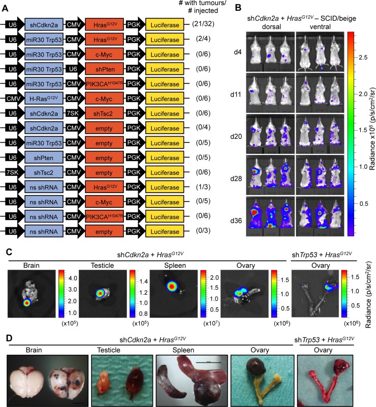
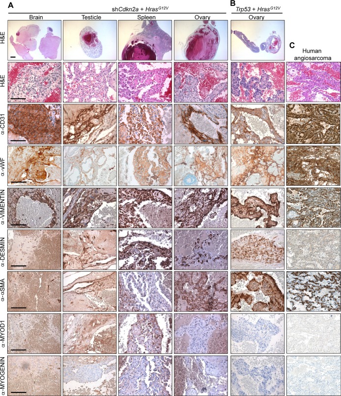
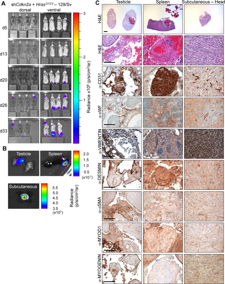
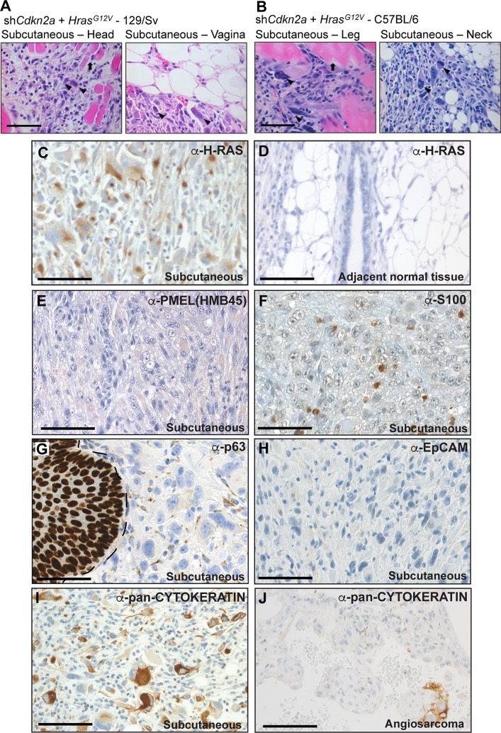
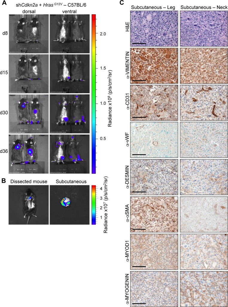
Soft tissue sarcomas are rare mesenchymal tumours accounting for 1% of adult malignancies and are fatal in approximately one third of patients. Two of the most aggressive and lethal forms of soft tissue sarcomas are angiosarcomas and undifferentiated pleomorphic sarcomas (UPS). To examine sarcoma-relevant molecular pathways, we employed a lentiviral gene regulatory system to attempt to generate in vivo models that reflect common molecular alterations of human angiosarcoma and UPS. Mice were intraveneously injected with MuLE lentiviruses expressing combinations of shRNA against Cdkn2a, Trp53, Tsc2 and Pten with or without expression of HrasG12V , PIK3CAH1047R or Myc. The systemic injection of an ecotropic lentivirus expressing oncogenic HrasG12V together with the knockdown of Cdkn2a or Trp53 was sufficient to initiate angiosarcoma and/or UPS development, providing a flexible system to generate autochthonous mouse models of these diseases. Unexpectedly, different mouse strains developed different types of sarcoma in response to identical genetic drivers, implicating genetic background as a contributor to the genesis and spectrum of sarcomas.

Keywords: H-Ras; MuLE lentivirus; angiosarcoma; mouse model; undifferentiated pleomorphic sarcoma.


Conflict of interest statement

CONFLICTS OF INTEREST The authors declare that they have no conflicts of interest related to this manuscript.

Figures

Figure 1
Figure 1. HrasG12V expression plus knockdown of Cdkn2a or Trp53 causes angiosarcomas in SCID/beige mice
(A) Schematic of MuLE vectors simultaneously expressing combinations of shRNAs against Cdkn2a, Trp53, Pten and Tsc2 with or without expression of HrasG12V, Myc or PIK3CAH1047R and a Luciferase reporter. (B) Bioluminescence imaging 4, 11, 20, 28 and 36 days after the injection of MuLE lentiviruses expressing shRNA against Cdkn2a together with HrasG12V into the tail veins of 4-6-week-old SCID/beige mice. Numbers of mice that developed tumours and total number of injected mice are shown for each construct. (C) Bioluminescence imaging and (D) photographs showing examples of tumour-bearing organs from shCdkn2a plus HrasG12V and shTrp53 plus HrasG12V injected animals. Scale bar: 10 mm.
Figure 2
Figure 2. Histological and molecular characterisation of angiosarcomas
H&E and immunohistochemical stainings using the indicated antibodies of tumours derived from the systemic injection of (A) shCdkn2a plus HrasG12V and (B) shTrp53 plus HrasG12V viruses. (C) Stainings of a human angiosarcoma tumour. Similar staining patterns were observed in two additional cases from different patients. Low magnification scale bars: 1000 μm and high magnification scale bars: 100 μm.
Figure 3
Figure 3. HrasG12V expression plus knockdown of Cdkn2a causes angiosarcomas and undifferentiated pleomorphic sarcomas in 129Sv mice
(A) Bioluminescence imaging 5, 13, 20, 26 and 33 days after the injection of MuLE lentiviruses expressing shRNA against Cdkn2a together with HrasG12V into the tail veins of 4-6-week-old 129/Sv mice. (B) Bioluminescence imaging showing examples of tumour-bearing organs.. (C) H&E and immunohistochemical stainings using the indicated antibodies. Low magnification scale bars: 1000 μm and high magnification scale bars: 100 μm.
Figure 4
Figure 4. High grade, locally invasive undifferentiated pleomorphic sarcomas form in 129/Sv and C57BL/6 mice
(A, B) High magnification images of H&E stained tumour sections derived from the intraveneous injection of shCdkn2a plus H-RasG12V viruses in (A) 129/Sv mice and (B) C57BL/6 mice. Arrowheads indicate atypical nuclei and arrows indicate entrapped muscle fibres. Images depict tumour invasion into the surrounding muscle or fat tissue. (C, D) Immunohistochemical staining using an anti-H-RAS antibody showing high H-RAS expression in a subcutaneous tumour (C) but absence of staining in adjacent normal tissue (D). (EI) Representative immunohistochemical staining patterns for the indicated antibodies in subcutaneous tumours. In (G) the margin between normal skin (p63 nuclear positive) and the tumour is shown by a dotted line. (J) Occasional cells in angiosarcoma tumours from SCID/beige mice stain positively for pan-CYTOKERATIN. Scale bars: 100 μm.
Figure 5
Figure 5. HrasG12V expression plus knockdown of Cdkn2a causes undifferentiated pleomorphic sarcomas in C57BL/6 mice
(A) Bioluminescence imaging 8, 15, 30 and 36 days after the injection of MuLE lentiviruses expressing shRNA against Cdkn2a together with HrasG12V into the tail veins of 4-6-week-old C57Bl/6 mice. (B) Bioluminescence imaging showing examples of a subcutaneous tumour prior to (left) and after (right) resection. (C) H&E and immunohistochemical stainings using the indicated antibodies. Scale bars: 100 μm.

References

    1. Fletcher C. WHO classification of tumours of soft tissue and bone. Fourth Edition. 2013 - PubMed
    1. Barretina J, Taylor BS, Banerji S, Ramos AH, Lagos-Quintana M, Decarolis PL, Shah K, Socci ND, Weir BA, Ho A, Chiang DY, Reva B, Mermel CH, et al. Subtype-specific genomic alterations define new targets for soft-tissue sarcoma therapy. Nat Genet. 2010;42:715–21. - PMC - PubMed
    1. Pérot G, Chibon F, Montero A, Lagarde P, de Thé H, Terrier P, Guillou L, Ranchère D, Coindre JM, Aurias A. Constant p53 Pathway Inactivation in a Large Series of Soft Tissue Sarcomas with Complex Genetics. The American Journal of Pathology. 2010;177:2080–90. - PMC - PubMed
    1. Borden EC, Baker LH, Bell RS, Bramwell V, Demetri GD, Eisenberg BL, Fletcher CD, Fletcher JA, Ladanyi M, Meltzer P, O'Sullivan B, Parkinson DR, Pisters PW, et al. Soft tissue sarcomas of adults: state of the translational science. Clin Cancer Res. 2003;9:1941–56. - PubMed
    1. Matushansky I, Charytonowicz E, Mills J, Siddiqi S, Hricik T, Cordon-Cardo C. MFH classification: differentiating undifferentiated pleomorphic sarcoma in the 21st Century. Expert Rev Anticancer Ther. 2009;9:1135–44. - PMC - PubMed