

Baicalein and baicalin inhibit colon cancer using two distinct fashions of apoptosis and senescence
- PMID: 29732005
- PMCID: PMC5929448
- DOI: 10.18632/oncotarget.24015
Baicalein and baicalin inhibit colon cancer using two distinct fashions of apoptosis and senescence
Abstract
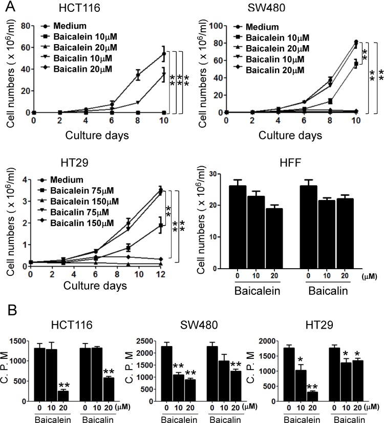
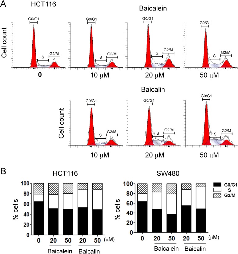
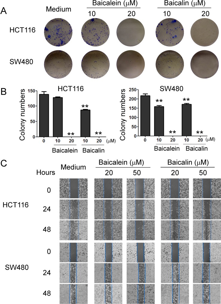
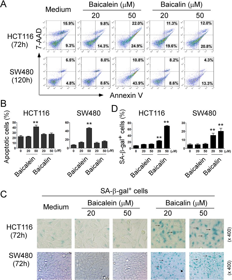
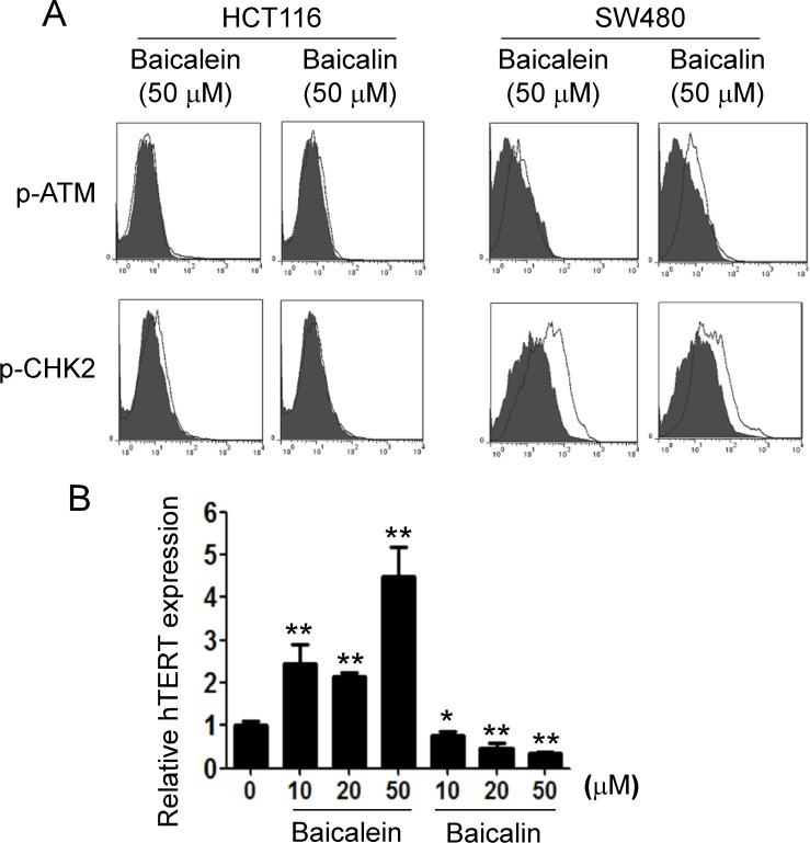
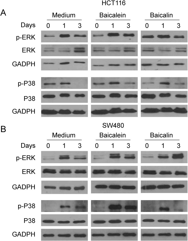
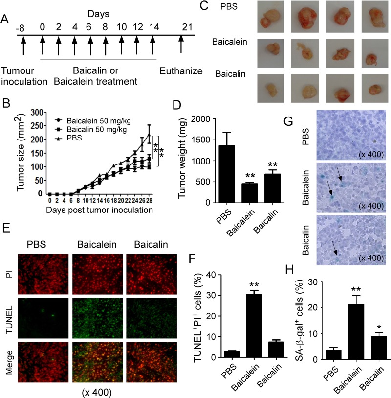
Baicalein and baicalin are active components of the Scutellaria baicalensis Georgi and both have broad anti-tumor activity. However, how and whether baicalein and baicalin inhibit colon cancer is unclear. Here we demonstrate that baicalein and baicalin can significantly inhibit human colon cancer cell growth and proliferation. Furthermore, both can induce cell cycle arrest, and suppress cancer cell colony formation and migration. The suppressive effects are mechanistically due to the induction of colon cancer cell apoptosis and senescence mediated by baicalein and baicalin, respectively. Furthermore, we revealed that baicalin-induced senescence in tumor cells is due to its inhibition of telomerase reverse transcriptase expression in tumor cells, and that MAPK ERK and p38 signaling pathways are causatively involved in the regulation of colon cancer cell apoptosis and senescence mediated by baicalein and baicalin. In addition, our in vivo studies using human colon cancer cells in humanized mouse xenograft models, further demonstrated that baicalein and baicalin can induce tumor cell apoptosis and senescence, resulting in inhibition of tumorigenesis and growth of colon cancer in vivo. These data clearly suggest that baicalein and baicalin have potent anti-cancer effects against human colon cancer and could be potential novel and effective target drugs for cancer therapy.
Keywords: Scutellaria baicalensis georgi; apoptosis; baicalein; baicalin; colon cancer.
Conflict of interest statement
CONFLICTS OF INTEREST The authors declare no competing financial interests.
Figures
Similar articles
-
Baicalein and Baicalin Promote Melanoma Apoptosis and Senescence via Metabolic Inhibition.Front Cell Dev Biol. 2020 Aug 25;8:836. doi: 10.3389/fcell.2020.00836. eCollection 2020. Front Cell Dev Biol. 2020. PMID: 32984331 Free PMC article.
-
Colon cancer chemopreventive effects of baicalein, an active enteric microbiome metabolite from baicalin.Int J Oncol. 2015 Nov;47(5):1749-58. doi: 10.3892/ijo.2015.3173. Epub 2015 Sep 18. Int J Oncol. 2015. PMID: 26398706 Free PMC article.
-
Baicalein, an active component of Scutellaria baicalensis Georgi, induces apoptosis in human colon cancer cells and prevents AOM/DSS-induced colon cancer in mice.Int J Oncol. 2013 Nov;43(5):1652-8. doi: 10.3892/ijo.2013.2086. Epub 2013 Sep 4. Int J Oncol. 2013. PMID: 24008356
-
Advances in the role of baicalin and baicalein in colon cancer: mechanisms and therapeutic potential.Discov Oncol. 2025 May 22;16(1):860. doi: 10.1007/s12672-025-02719-5. Discov Oncol. 2025. PMID: 40404880 Free PMC article. Review.
-
An overview of pharmacological activities of baicalin and its aglycone baicalein: New insights into molecular mechanisms and signaling pathways.Iran J Basic Med Sci. 2022 Jan;25(1):14-26. doi: 10.22038/IJBMS.2022.60380.13381. Iran J Basic Med Sci. 2022. PMID: 35656442 Free PMC article. Review.
Cited by
-
Anticancer activities of phytoconstituents and their liposomal targeting strategies against tumor cells and the microenvironment.Adv Drug Deliv Rev. 2020;154-155:245-273. doi: 10.1016/j.addr.2020.05.006. Epub 2020 May 28. Adv Drug Deliv Rev. 2020. PMID: 32473991 Free PMC article. Review.
-
Unwinding Helicase MCM Functionality for Diagnosis and Therapeutics of Replication Abnormalities Associated with Cancer: A Review.Mol Diagn Ther. 2024 May;28(3):249-264. doi: 10.1007/s40291-024-00701-5. Epub 2024 Mar 26. Mol Diagn Ther. 2024. PMID: 38530633 Review.
-
Incredible use of plant-derived bioactives as anticancer agents.RSC Adv. 2025 Jan 20;15(3):1721-1746. doi: 10.1039/d4ra05089d. eCollection 2025 Jan 16. RSC Adv. 2025. PMID: 39835210 Free PMC article. Review.
-
Interplay Between Traditional and Scientific Knowledge: Phytoconstituents and Their Roles in Lung and Colorectal Cancer Signaling Pathways.Biomolecules. 2025 Mar 5;15(3):380. doi: 10.3390/biom15030380. Biomolecules. 2025. PMID: 40149916 Free PMC article. Review.
-
Therapeutic efficacy of baicalein and 6-(methylsulfinyl)hexyl isothiocyanate, alone or in combination with 5-fluorouracil, in the treatment of colorectal cancer.Naunyn Schmiedebergs Arch Pharmacol. 2025 Jun 10. doi: 10.1007/s00210-025-04274-w. Online ahead of print. Naunyn Schmiedebergs Arch Pharmacol. 2025. PMID: 40493247
References
-
- Siegel R, Naishadham D, Jemal A. Cancer statistics, 2012. CA Cancer J Clin. 2012;62:10–29. https://doi.org/10.3322/caac.20138 - DOI - PubMed
-
- Siegel R, Naishadham D, Jemal A. Cancer statistics, 2013. CA Cancer J Clin. 2013;63:11–30. https://doi.org/10.3322/caac.21166 - DOI - PubMed
-
- Siegel RL, Miller KD, Jemal A. Cancer statistics, 2015. CA Cancer J Clin. 2015;65:5–29. https://doi.org/10.3322/caac.21254 - DOI - PubMed
-
- Panczyk M. Pharmacogenetics research on chemotherapy resistance in colorectal cancer over the last 20 years. World J Gastroenterol. 2014;20:9775–827. https://doi.org/10.3748/wjg.v20.i29.9775 - DOI - PMC - PubMed
-
- Chia JS, Du JL, Hsu WB, Sun A, Chiang CP, Wang WB. Inhibition of metastasis, angiogenesis, and tumor growth by Chinese herbal cocktail Tien-Hsien Liquid. BMC Cancer. 2010;10:175. https://doi.org/10.1186/1471-2407-10-175 - DOI - PMC - PubMed
LinkOut - more resources
Full Text Sources
Other Literature Sources
Miscellaneous
