

Granular cell tumour of the neurohypophysis: an unusual cause of hypopituitarism
- PMID: 29732160
- PMCID: PMC5931226
- DOI: 10.1530/EDM-17-0178
Granular cell tumour of the neurohypophysis: an unusual cause of hypopituitarism
Abstract
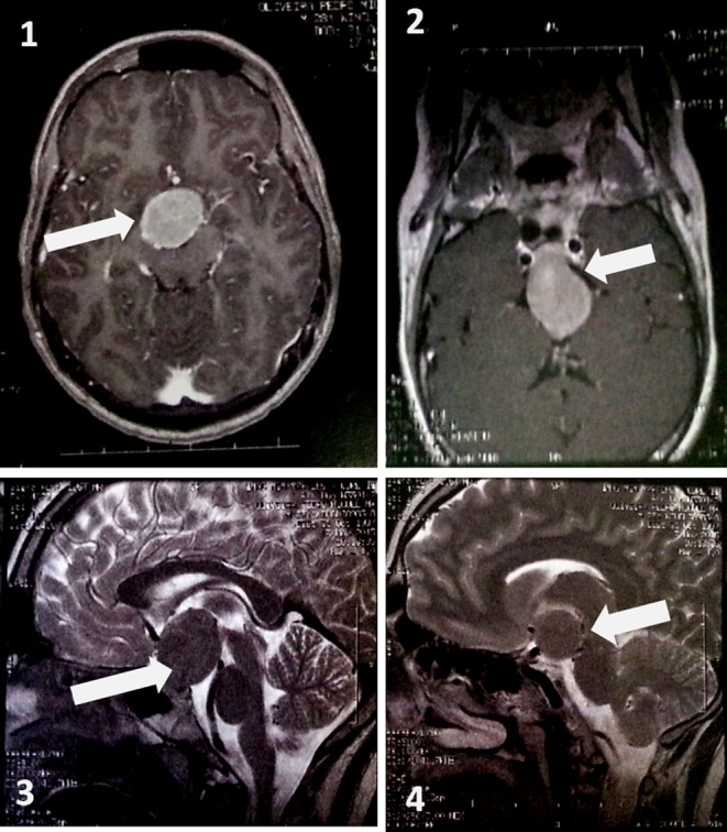
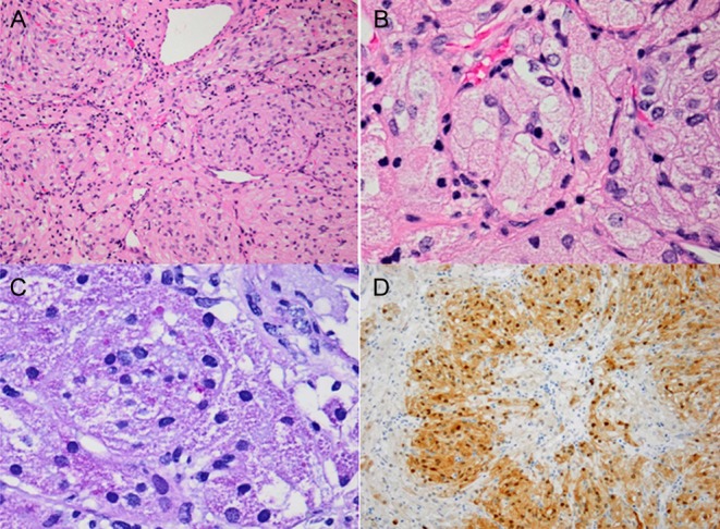
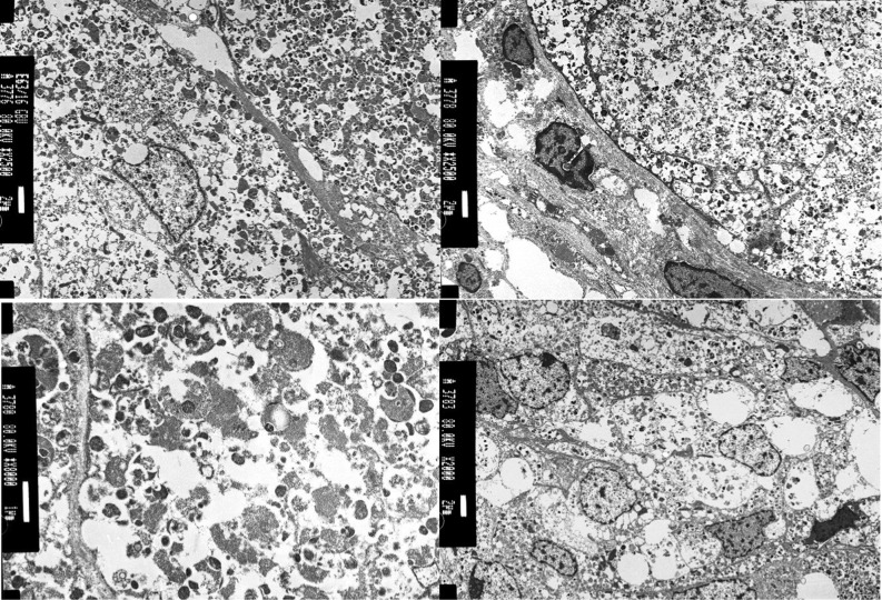
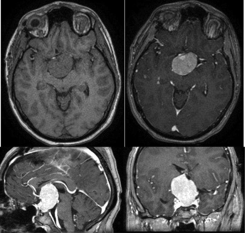
Granular cell tumours (GCT) are rare, slow-growing, benign neoplasms that are usually located in the head and neck. They are more frequent in the female gender and typically have an asymptomatic clinical course, being diagnosed only at autopsy. Symptomatic GCT of the neurohypophysis are exceedingly rare, being less than 70 cases described so far. The authors report on a case of a 28-year-old male that presented to the Endocrinology clinic with clinical and biochemical evidence of hypogonadism. He also reported minor headaches without any major visual symptoms. Further laboratory tests confirmed hypopituitarism (hypogonadotrophic hypogonadism, central hypothyroidism and hypocortisolism) and central nervous system imaging revealed a pituitary macroadenoma. The patient underwent transcranial pituitary adenoma resection and the pathology report described a GCT of the neurohypophysis with low mitotic index. The reported case is noteworthy for the rarity of the clinicopathological entity.
Learning points: Symptomatic GCTs are rare CNS tumours whose cell of origin is not well defined that usually give rise to visual symptoms, headache and endocrine dysfunction.Imaging is quite unspecific and diagnosis is difficult to establish preoperatively.Surgical excision is challenging due to lesion's high vascularity and propensity to adhere to adjacent structures.The reported case is noteworthy for the rarity of the clinicopathological entity.
Figures
References
-
- Ostrom QT, Gittleman H, Fulop J, Liu M, Blanda R, Kromer C, Wolinsky Y, Kruchko C, Barnholtz-Sloan JS. CBTRUS Statistical Report: primary brain and central nervous system tumors diagnosed in the United States in 2008–2012. Neuro-Oncology 2015. 17 (Supplement 4) iv1 (10.1093/neuonc/nov189) - DOI - PMC - PubMed
-
- Latini F, Ambrosio MR, Guerra A, Uberti ED, Cavallo MA, Lapparelli M. Pituitary granular cell tumor: single-center experience and comprehensive update. Contemporary Neurosurgery 2014. 36 1–7. (10.1097/01.CNE.0000455825.70290.92) - DOI
-
- Boyce R, Beadles CF. A further contribution to the study of the pathology of the hypophysis cerebri. Journal of Pathology and Bacteriology 1893. 1 359–383. (10.1002/path.1700010310) - DOI
LinkOut - more resources
Full Text Sources
Other Literature Sources
