

Changes in buccal facial depth of female patients after extraction and nonextraction orthodontic treatments: A preliminary study
- PMID: 29732303
- PMCID: PMC5932319
- DOI: 10.4041/kjod.2018.48.3.172
Changes in buccal facial depth of female patients after extraction and nonextraction orthodontic treatments: A preliminary study
Abstract
Objective: This study was performed to investigate buccal facial depth (BFD) changes after extraction and nonextraction orthodontic treatments in post-adolescent and adult female patients, and to explore possible influencing factors.
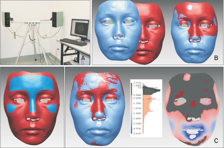
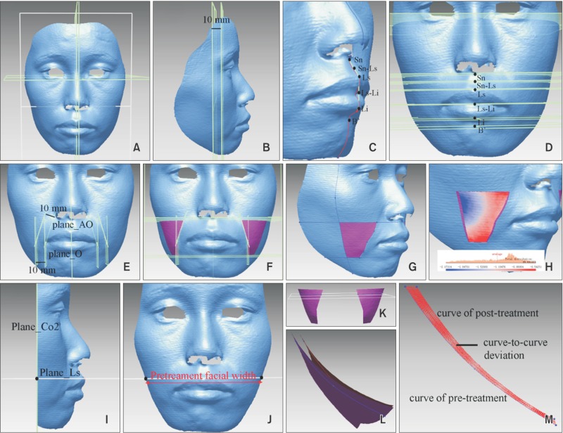
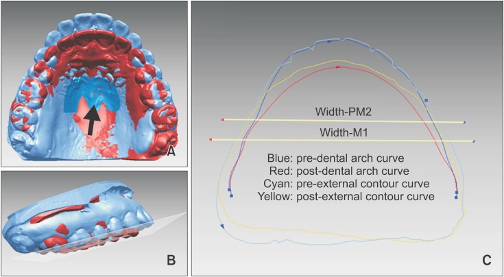
Methods: Twelve and nine female patients were enrolled in the extraction and nonextraction groups, respectively. Changes in BFD in the defined buccal region and six transverse and two coronal measuring planes were measured after registering pretreatment and posttreatment three-dimensional facial scans. Changes in posterior dentoalveolar arch widths were also measured. Treatment duration, changes in body mass index (BMI), and cephalometric variables were compared between the groups.
Results: BFD in the buccal region decreased by approximately 1.45 mm in the extraction group, but no significant change was observed in the nonextraction group. In the extraction group, the decrease in BFD was identical between the two coronal measuring planes, whereas this differed among the six transverse measuring planes. Posterior dentoalveolar arch widths decreased in the extraction group, whereas these increased at the second premolar level in the nonextraction group. The treatment duration of the extraction group was twice that of the nonextraction group. No differences were found in BMI and Frankfort horizontal-mandibular plane angle changes between the groups. BFD changes in the buccal region moderately correlated with treatment duration and dental arch width change.
Conclusions: BFD decreased in adult female patients undergoing extraction, and this may be influenced by the long treatment duration and constriction of dentoalveolar arch width. However, nonextraction treatment did not significantly alter BFD.
Keywords: Adult treatment; Extraction vs. nonextraction; Soft tissue; Three-dimensional scanner.
Conflict of interest statement
CONFLICTS OF INTEREST: No potential conflict of interest relevant to this article was reported.
Figures
References
-
- Luppanapornlarp S, Johnston LE., Jr The effects of premolar-extraction: a long-term comparison of outcomes in “clear-cut” extraction and nonextraction Class II patients. Angle Orthod. 1993;63:257–272. - PubMed
-
- Scott SH, Johnston LE., Jr The perceived impact of extraction and nonextraction treatments on matched samples of African American patients. Am J Orthod Dentofacial Orthop. 1999;116:352–360. - PubMed
-
- Bowman SJ, Johnston LE., Jr The esthetic impact of extraction and nonextraction treatments on Caucasian patients. Angle Orthod. 2000;70:3–10. - PubMed
-
- Basciftci FA, Usumez S. Effects of extraction and nonextraction treatment on class I and class II subjects. Angle Orthod. 2003;73:36–42. - PubMed
-
- Wholley CJ, Woods MG. The effects of commonly prescribed premolar extraction sequences on the curvature of the upper and lower lips. Angle Orthod. 2003;73:386–395. - PubMed
LinkOut - more resources
Full Text Sources
Other Literature Sources
Miscellaneous