

Evaluation of the Effect of Source Geometry on the Output of Miniature X-ray Tube for Electronic Brachytherapy through Simulation
- PMID: 29732338
- PMCID: PMC5928308
Evaluation of the Effect of Source Geometry on the Output of Miniature X-ray Tube for Electronic Brachytherapy through Simulation
Abstract
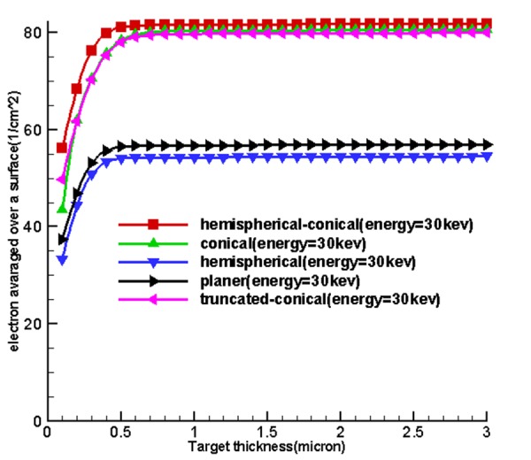
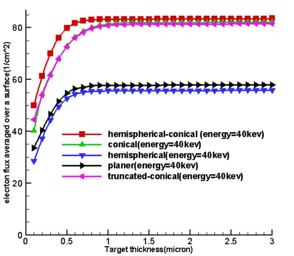
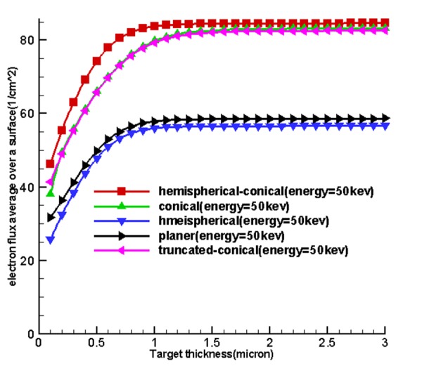
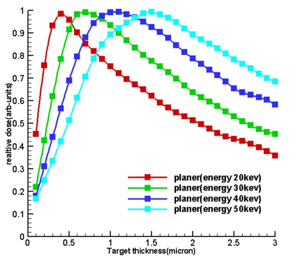
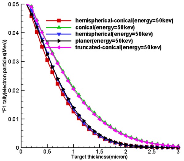
Objective: The use of miniature X-ray source in electronic brachytherapy is on the rise so there is an urgent need to acquire more knowledge on X-ray spectrum production and distribution by a dose. The aim of this research was to investigate the influence of target thickness and geometry at the source of miniature X-ray tube on tube output.
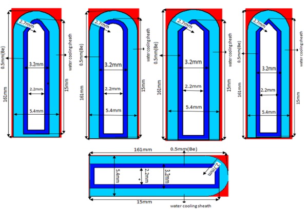
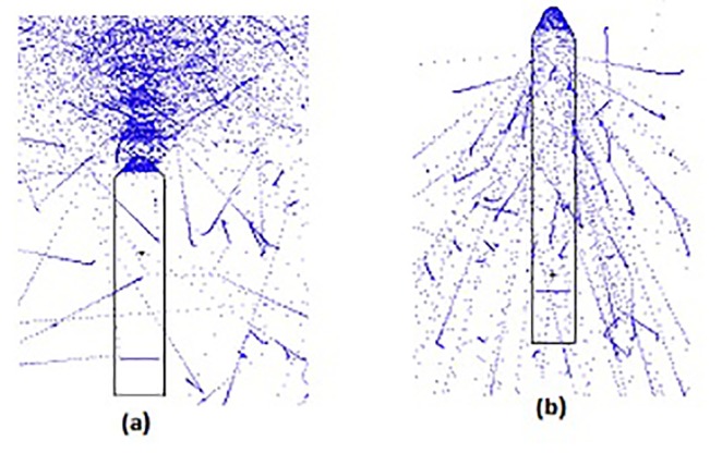
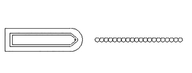
Method: Five sources were simulated based on problems each with a specific geometric structure and conditions using MCNPX code. Tallies proportional to the output were used to calculate the results for the influence of source geometry on output.
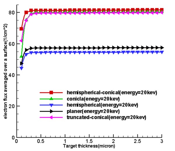
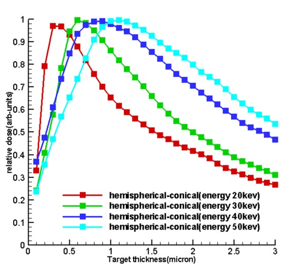
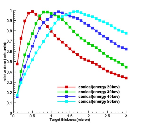
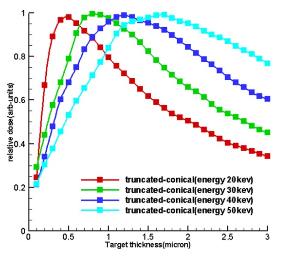
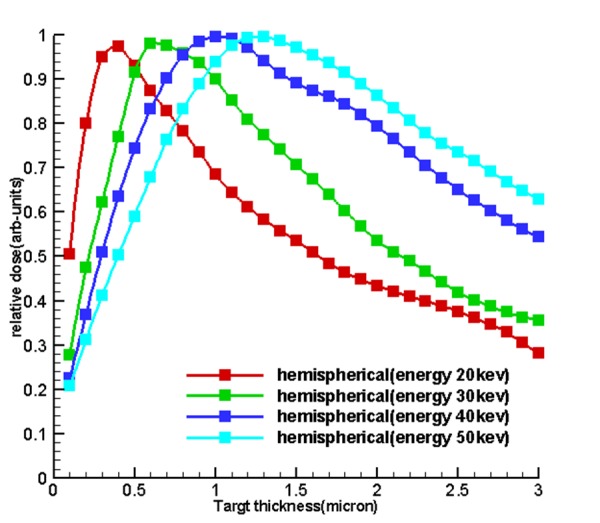
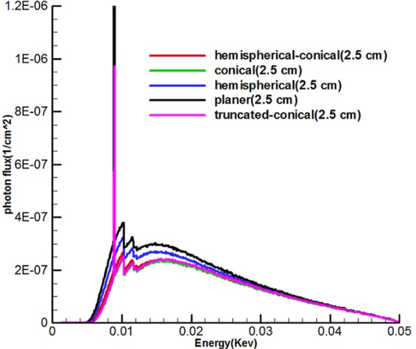
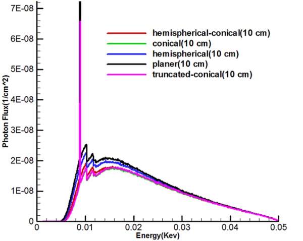
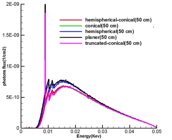
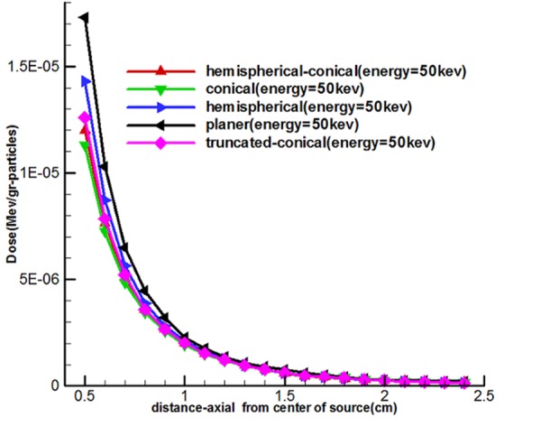
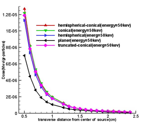
Results: The results of this work include the size of the optimal thickness of 5 miniature sources, energy spectrum of the sources per 50 kev and also the axial and transverse dose of simulated sources were calculated based on these thicknesses. The miniature source geometric was affected on the output x-ray tube.
Conclusion: The result of this study demonstrates that hemispherical-conical, hemispherical and truncated-conical miniature sources were determined as the most suitable tools.
Keywords: Electronic Brachytherapy; Energy Spectrum; Miniature Source; Target Optimization; Monte Carlo.
Figures
Similar articles
-
Assessment of two hemispherical and hemispherical-conical miniature sources used in electronic brachytherapy using Monte Carlo Simulation.Electron Physician. 2017 Feb 25;9(2):3845-3856. doi: 10.19082/3845. eCollection 2017 Feb. Electron Physician. 2017. PMID: 28465817 Free PMC article.
-
Characteristics of miniature electronic brachytherapy x-ray sources based on TG-43U1 formalism using Monte Carlo simulation techniques.Med Phys. 2012 Apr;39(4):1971-9. doi: 10.1118/1.3693046. Med Phys. 2012. PMID: 22482618
-
Anode optimization for miniature electronic brachytherapy X-ray sources using Monte Carlo and computational fluid dynamic codes.J Adv Res. 2016 Mar;7(2):225-32. doi: 10.1016/j.jare.2015.04.006. Epub 2015 Apr 20. J Adv Res. 2016. PMID: 26966563 Free PMC article.
-
Simulation evaluation of NIST air-kerma rate calibration standard for electronic brachytherapy.Med Phys. 2016 Mar;43(3):1119-29. doi: 10.1118/1.4940791. Med Phys. 2016. PMID: 26936699
-
A Monte Carlo-based dosimetric characterization of Esteya® , an electronic surface brachytherapy unit.Med Phys. 2019 Jan;46(1):356-369. doi: 10.1002/mp.13275. Epub 2018 Nov 28. Med Phys. 2019. PMID: 30390317
References
-
- Cho SO, Heo SH. Super miniature X-ray tube using NANO material field emitter. United States: Google Patents; 2012.
LinkOut - more resources
Full Text Sources