Review
. 2018 Aug;7(15):e1701461.
doi: 10.1002/adhm.201701461. Epub 2018 May 7.

Tissue Engineering at the Blood-Contacting Surface: A Review of Challenges and Strategies in Vascular Graft Development

Affiliations
Review

Tissue Engineering at the Blood-Contacting Surface: A Review of Challenges and Strategies in Vascular Graft Development

Daniel Radke et al. Adv Healthc Mater. 2018 Aug.

Abstract

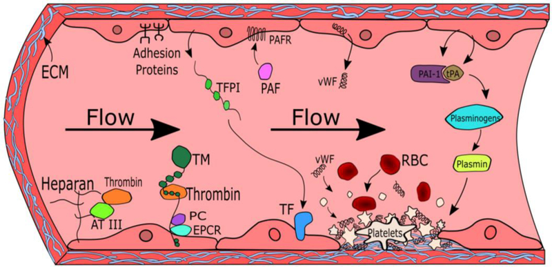
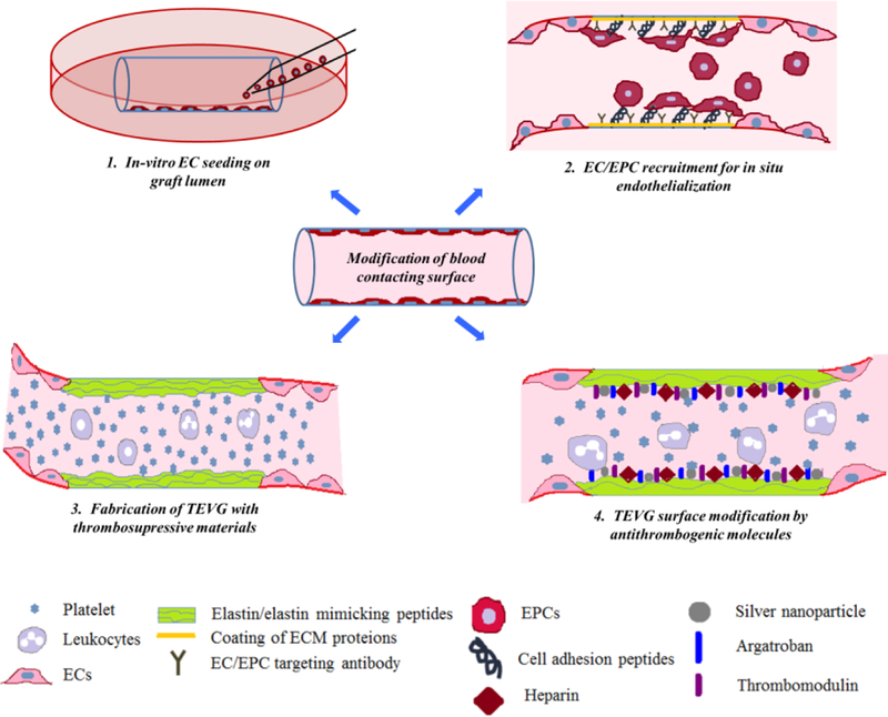
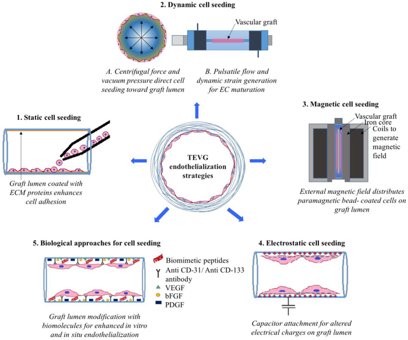
Tissue engineered vascular grafts (TEVGs) are beginning to achieve clinical success and hold promise as a source of grafting material when donor grafts are unsuitable or unavailable. Significant technological advances have generated small-diameter TEVGs that are mechanically stable and promote functional remodeling by regenerating host cells. However, developing a biocompatible blood-contacting surface remains a major challenge. The TEVG luminal surface must avoid negative inflammatory responses and thrombogenesis immediately upon implantation and promote endothelialization. The surface has therefore become a primary focus for research and development efforts. The current state of TEVGs is herein reviewed with an emphasis on the blood-contacting surface. General vascular physiology and developmental challenges and strategies are briefly described, followed by an overview of the materials currently employed in TEVGs. The use of biodegradable materials and stem cells requires careful control of graft composition, degradation behavior, and cell recruitment ability to ensure that a physiologically relevant vessel structure is ultimately achieved. The establishment of a stable monolayer of endothelial cells and the quiescence of smooth muscle cells are critical to the maintenance of patency. Several strategies to modify blood-contacting surfaces to resist thrombosis and control cellular recruitment are reviewed, including coatings of biomimetic peptides and heparin.

Keywords: blood-contacting surfaces; endothelialization; surface modification; vascular engineering; vascular grafts.


Figures

Figure 1.
Figure 1.. The role of endothelial cells (ECs) in the regulation of thrombogenesis.
Heparans, thrombomodulin, tissue plasminogen activator (tPA), plasminogen activator inhibitor-1 (PAI-1), adhesion proteins, and platelet activating factor (PAF) play key roles in regulating coagulation. These important factors interact with other molecules such as thrombin, antithrombin III (AT III), protein C, endothelial protein coupled receptor (EPCR), platelet activating factor receptor (PAFR), plasmin, and plasminogen to promote or prevent thrombus formation. Thrombi are formed when damaged endothelium expose extracellular matrix (ECM) proteins such as collagen, which then recruit platelets, von Willebrand factor (vWF), and red blood cells (RBCs).
Figure 2.
Figure 2.. Modifying blood-contacting surface of TEVG.
(1). In vitro endothelial cell seeding on the graft lumen provides a biomimetic environment and prevents direct contact of blood with the graft surface. (2). In-situ endothelialization can be achieved by coating the graft lumen with ECM proteins, growth factors, EC/EPC specific cell binding peptides or antibodies. (3). Fabrication of TEVG with anti-thrombogenic natural or synthetic materials maintains graft patency by preventing platelet adhesion and activation. (4). Coating of various anti-thrombogenic molecules such as heparin, silver nano-particles, argatroban and thrombomodulin actively maintains graft patency by preventing platelet adhesion and activation.
Figure 3:
Figure 3:. Cell-seeding strategies for TEVG Endothelialization.
(1). Static cell seeding: ECs/ EPCs can be directly seeded on the vascular graft lumen. A graft lumen can be coated with individual or combinations of various ECM proteins for enhanced cell attachment and retention [144, 145]. (2). Dynamic cell seeding: (A) Vacuum pressure and centrifugal force applied toward graft lumen increases cell seeding efficiency and reduces cell culture period [146, 147]. (B) Bioreactors can provide a physiological pulsatile flow environment, which can be used to promote a mature, non-activated EC phenotype [–150]. (3). Magnetic cell seeding: An external magnetic field can efficiently guide and regulate seeding of paramagnetic/superparamagnetic particle coated EC/EPCs on a graft lumen [–153]. (4). Electrostatic cell seeding: attachment of e-PTFE graft with capacitor generates temporary positive charge on graft lumen. Cell surface is usually negatively charged; thus, this temporary positive charge on graft lumen can be used for successful adhesion of ECs/EPCs [154]. (5). Biological approaches for cell seeding: A graft lumen can be coated with biomimetic cell adhesion peptides [–164], specific antibodies to capture ECs/EPCs [–168] and various growth factors to promote in vitro/in situ migration and maturation of ECs/EPCs during cell culture and/or after implantation [–173].
Figure 4.
Figure 4.. Inclusion of anticoagulation molecules in TEVGs.
(1) Thrombomodulin (TM) is a transmembrane protein present on ECs that binds thrombin. The thrombin-TM complex activates protein C (PC, APC), which then forms a membrane-bound complex with protein S (S) that inactivates factors Va and VIIIa. Recombinant TM can be covalently bound to synthetic materials [209]. (2) Argatroban locally and directly inhibits thrombin by non-covalently interacting with Asp-189 in the S1 binding pocket of thrombin to block its enzymatic activity. Truncated forms were developed which retained the guanidyl group for ionic interaction and featured a spacer arm on the piperazinyl amide moiety for surface grafting [210]. (3) Nitric oxide (NO) activates soluble guanylate cyclase (sGC) to increase cyclic guanosine monophosphate (cGMP) production by binding to the sixth position of the heme ring, breaking the bond to His-105. cGMP then decreases expression of thromboxane A2 and the platelet surface adhesion protein P-selectin. (4) Silver nanoparticles (SNP) inhibit platelet aggregation by blocking biochemical pathways associated with membrane restructuring, preventing the expression of surface adhesion proteins.

References

    1. Xu J, Murphy SL, Kochanek KD, Bastian BA, National Vital Statistics Reports 2016, 64, 39. - PubMed
    1. Mendis S, et al., The World Health Organization, Geneva, Switzerland: 2014.
    1. Pashneh-Tala S, MacNeil S, Claeyssens F, Tissue Engineering. Part B, Reviews 2016, 22, 68. - PMC - PubMed
    1. Lin S, Sandig M, Mequanint K, Tissue engineering. Part A 2011, 17, 1561. - PubMed
    1. Rubanyi GM, J Cardiovasc Pharmacol 1993, 22 Suppl 4, S1. - PubMed

Publication types