

Ex Utero Electroporation and Organotypic Slice Cultures of Embryonic Mouse Brains for Live-Imaging of Migrating GABAergic Interneurons
- PMID: 29733310
- PMCID: PMC6100702
- DOI: 10.3791/57526
Ex Utero Electroporation and Organotypic Slice Cultures of Embryonic Mouse Brains for Live-Imaging of Migrating GABAergic Interneurons
Abstract
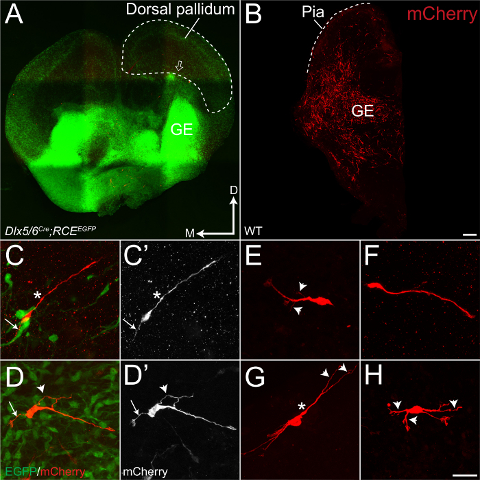
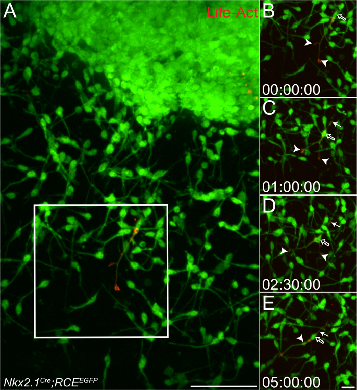
GABAergic interneurons (INs) are critical components of neuronal networks that drive cognition and behavior. INs destined to populate the cortex migrate tangentially from their place of origin in the ventral telencephalon (including from the medial and caudal ganglionic eminences (MGE, CGE)) to the dorsal cortical plate in response to a variety of intrinsic and extrinsic cues. Different methodologies have been developed over the years to genetically manipulate specific pathways and investigate how they regulate the dynamic cytoskeletal changes required for proper IN migration. In utero electroporation has been extensively used to study the effect of gene repression or overexpression in specific IN subtypes while assessing the impact on morphology and final position. However, while this approach is readily used to modify radially migrating pyramidal cells, it is more technically challenging when targeting INs. In utero electroporation generates a low yield given the decreased survival rates of pups when electroporation is conducted before e14.5, as is customary when studying MGE-derived INs. In an alternative approach, MGE explants provide easy access to the MGE and facilitate the imaging of genetically modified INs. However, in these explants, INs migrate into an artificial matrix, devoid of endogenous guidance cues and thalamic inputs. This prompted us to optimize a method where INs can migrate in a more naturalistic environment, while circumventing the technical challenges of in utero approaches. In this paper, we describe the combination of ex utero electroporation of embryonic mouse brains followed by organotypic slice cultures to readily track, image and reconstruct genetically modified INs migrating along their natural paths in response to endogenous cues. This approach allows for both the quantification of the dynamic aspects of IN migration with time-lapse confocal imaging, as well as the detailed analysis of various morphological parameters using neuronal reconstructions on fixed immunolabeled tissue.


References
Publication types
MeSH terms
Grants and funding
LinkOut - more resources
Full Text Sources
Other Literature Sources
Medical
Research Materials
