. 2019 Feb 1;16(2):217-225.
doi: 10.1093/ons/opy083.

Minimally Invasive Bilateral Anterior Cingulotomy via Open Minicraniotomy Using a Novel Multiport Cisternoscope: A Cadaveric Demonstration

Affiliations

Minimally Invasive Bilateral Anterior Cingulotomy via Open Minicraniotomy Using a Novel Multiport Cisternoscope: A Cadaveric Demonstration

Sunil Manjila et al. Oper Neurosurg. .

Abstract

Background: Bilateral anterior cingulotomy has been used to treat chronic pain, obsessive compulsive disorder, and addictions. Lesioning of the target area is typically performed using bilateral stereotactic electrode placement and target ablation, which involves transparenchymal access through both hemispheres.

Objective: To evaluate an endoscopic direct-vision lesioning using a unilateral parasagittal minicraniotomy for minimally invasive bilateral anterior cingulotomy using a novel multiport endoscope through the anterior interhemispheric fissure.

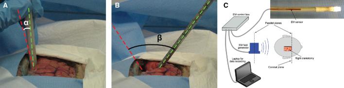
Methods: A novel multiport magnetic resonance imaging (MRI)-compatible neuroendoscope prototype is used to demonstrate cadaveric cingulate lesioning through a lateral imaging port while simultaneously viewing the pericallosal arteries as landmarks through a tip imaging port. The lateral port enables extended lesioning of the gyrus while rotation of the endoscope about its axis provides access to homologous areas of both hemispheres.

Results: Cadaver testing confirmed the capability to navigate the multiport neuroendoscope between the hemispheres using concurrent imaging from the tip and lateral ports. The lateral port enabled exploration of the gyrus, visualization of lesioning, and subsequent inspection of lesions. Tip-port imaging provided navigational cues and allowed the operator to ensure that the endoscope tip did not contact tissue. The multiport design required instrument rotation in the coronal plane of only 20° to lesion both gyri, while a standard endoscope necessitated a rotation of 54°.

Conclusion: Multiport MRI-compatible endoscopy can be effectively used in cisternal endoscopy, whereby a unilateral parasagittal minicraniotomy can be used for endoscopic interhemispheric bilateral anterior cingulotomy.


Figures

FIGURE 1.
FIGURE 1.
MRI scan images after cingulotomy showing 3 classic ablation sites in the sagittal plane adjacent to each other, A, also revealing the subcallosal gyrus (white asterisk) as a preferred additional lesioning target, which also can be accessed interhemipsherically. A and B depict the lines of insertion of ablation electrodes through cranial burr holes, while C depicts the proposed interhemispheric approach via open minicraniotomy using a multiport endoscope between the bridging veins. Note the retraction-free axial rotation of the cisternal endoscope in such a way that the side port faces the treated ACCs by 180 ° turns. © 2014 JNSPG. Reprinted with permission from Yang et al, J Neurosurg.
FIGURE 2.
FIGURE 2.
Schematic of unilateral minicraniotomy and interhemispheric approach to bilateral cingulate gyri using neuronendoscope incorporating 2 imaging and instrument ports. The tip port is used to guide endoscopic instrument insertion and navigation while the lateral port is used for lesion creation and inspection. Both gyri can be accessed via rotation of the endoscope about its axis.
FIGURE 3.
FIGURE 3.
Computer aided design and photograph of multiport neuroendoscope prototype. The front port enables visualization of the pericallosal arteries while the side port is used for lesioning of both cingulate gyri.
FIGURE 4.
FIGURE 4.
Technique for retractor-free access into the interhemispheric cistern. A, Inflatable flat-balloon catheter designed for interhemispheric soft brain retraction. B, Gradual inflation of the catheter in the interhemispheric cistern to avoid use of metal retractors and resultant direct impact brain injury. C, Multiport neuroendoscope entering the cistern between draining veins. In B and C, locations of parasagittal draining veins are marked with dashed black lines across the interhemispheric fissure. Note that depicted craniotomy has been made large for visualization purposes.
FIGURE 5.
FIGURE 5.
Comparison of coronal-plane pivoting angles (α and β) necessary to create lesions in both hemispheres using A, multiport prototype and B, standard neuroendoscope. Red line indicates instrument position for ipsilateral cingulotomy and green line represents contralateral instrument position. C, Electromagnetic measurement system (Trakstar, Ascension Technology, Shelburne, Vermont).
FIGURE 6.
FIGURE 6.
Landmark-based endoscope navigation of the interhemispheric fissure using the front viewing port with 3 transparent working channels. A, Initial visualization of supracallosal vessels after parafalcine insertion of multiport endoscope. B, View showing the parallel-running pericallosal arteries. C, View showing localization of the CMA in relation to PCA. D, Close-up view identifying the cingulate gyrus, marked C in between PCA and CMA. F: falx cerebri; PCA: pericallosal artery, CMA: callosomarginal artery.
FIGURE 7.
FIGURE 7.
Side port views showing sequential positioning of Bugbee wire on opposing ACCs used to measure coronal-plane pivoting. A, Verification of the boundaries of cingulate gyrus by visualizing long axis of CMA. B, Contact with left ACC. C, Contact with right ACC after an axial turn of 180 °. D, View of ablation site on the right ACC.
FIGURE 8.
FIGURE 8.
A, Image mosaic demonstrating interhemispheric exploration of the left ACC using side port, anterior to posterior direction. Left callosomarginal artery can be seen curving upward at right side of image. B, Image mosaic of right ACC illustrating inspection of 3 lesions (arrows). Note that both arterial and venous contrast silicone dye did not adequately penetrate this hemisphere unlike the left hemisphere.

References

    1. Manjila S, Mencattelli M, Rosa B, Price K, Fagogenis G, Dupont PE. A multiport MR-compatible neuroendoscope: spanning the gap between rigid and flexible scopes. Neurosurg Focus. 2016;41(3):E13. - PMC - PubMed
    1. Ramesh V, Balasubraminian C. Letter to the editor: obsessive-compulsive disorder and cingulotomy. J Neurosurg. 2013;119(2):526. - PubMed
    1. Banks GP, Mikell CB, Youngerman BEet al. Neuroanatomical characteristics associated with response to dorsal anterior cingulotomy for obsessive-compulsive disorder. JAMA Psychiatry. 2015;72(2):127-135. - PubMed
    1. Bourne SK, Sheth SA, Neal Jet al. Beneficial effect of subsequent lesion procedures after nonresponse to initial cingulotomy for severe, treatment-refractory obsessive-compulsive disorder. Neurosurgery. 2013;72(2):196-202. - PubMed
    1. De Ridder D, Leong SL, Manning P, Vanneste S, Glue P. Anterior cingulate implant for obsessive-compulsive disorder. World Neurosurg. 2017;97:754.e7-754.e16. - PubMed

Publication types