Comparative Study
. 2018 Oct 1;198(7):880-890.
doi: 10.1164/rccm.201712-2423OC.

Blood Neutrophils Are Reprogrammed in Bronchiectasis

Affiliations
Comparative Study

Blood Neutrophils Are Reprogrammed in Bronchiectasis

Pallavi Bedi et al. Am J Respir Crit Care Med. .

Abstract

Rationale: Excessive neutrophilic airway inflammation is the central feature of bronchiectasis, but little is known about neutrophils in bronchiectasis.

Objectives: To assess blood neutrophil phenotype in patients with bronchiectasis while stable and during exacerbations.

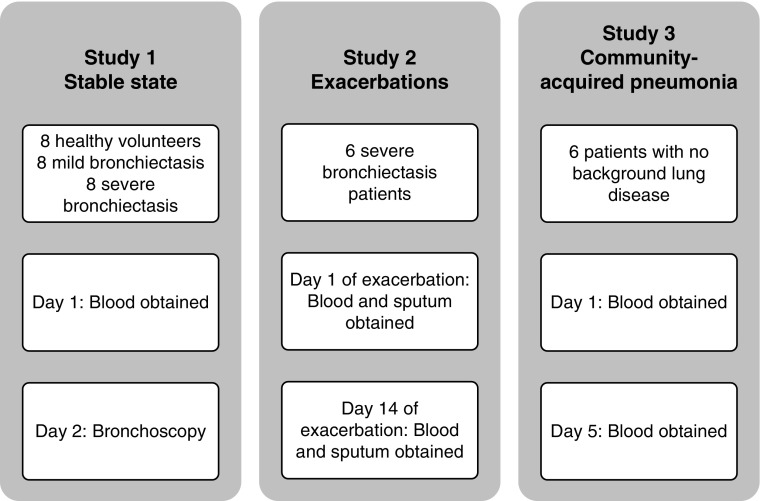
Methods: In the clinically stable arm of this study, there were eight healthy volunteers, eight patients with mild bronchiectasis, and eight patients with severe bronchiectasis. In addition, six patients with severe bronchiectasis were compared with six patients with community-acquired pneumonia at the start and end of an exacerbation. We assessed neutrophils for spontaneous apoptosis, cell surface marker expression, degranulation, reactive oxygen species generation, phagocytosis, and killing of Pseudomonas aeruginosa (PAO1). In addition, blood neutrophil function was compared with airway neutrophil function in bronchiectasis.

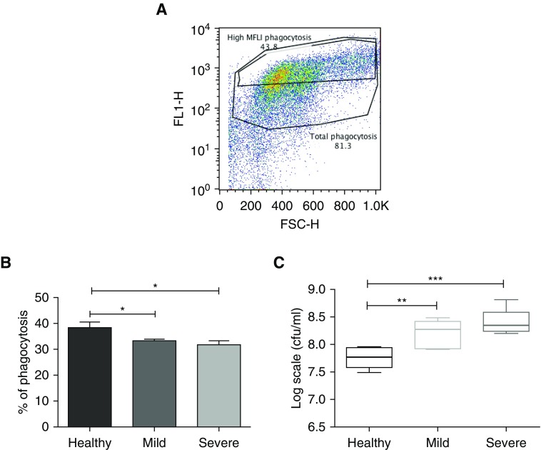
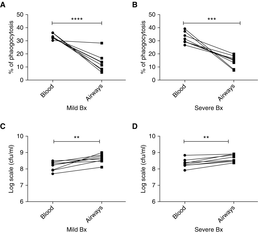
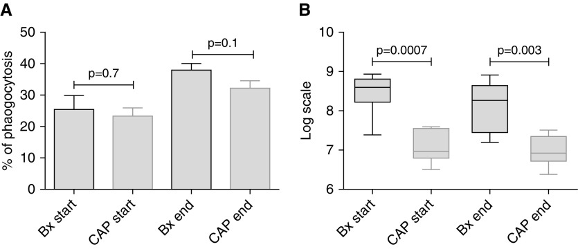
Measurements and main results: In stable bronchiectasis, compared with healthy volunteers, blood neutrophils had significantly prolonged viability, delayed apoptosis, increased CD62L shedding, upregulated CD11b expression, increased myeloperoxidase release, and impaired neutrophil phagocytosis and killing of PAO1. Bronchiectatic airway neutrophils had significantly lower bacterial phagocytosis and killing than their matched autologous blood neutrophils. Both blood and airway neutrophil phagocytosis and killing were impaired at the start of an exacerbation and improved following antibiotic treatment. In pneumonia, there was a significant improvement in phagocytosis and killing after treatment with antibiotics. During infections, there was no difference in phagocytosis, but there was significantly increased bacterial killing at the start and end of infection in pneumonia compared with bronchiectasis exacerbations.

Conclusions: In bronchiectasis stable state, peripheral blood neutrophils are reprogrammed and have prolonged survival. This impairs their functional ability of bacterial phagocytosis and killing, thereby perpetuating the vicious circle in bronchiectasis.

Keywords: bronchiectasis; infection; inflammation; neutrophils.


Figures

Figure 1.
Figure 1.
The three groups recruited for this study. Study 1: all study participants (healthy volunteers, volunteers with mild bronchiectasis, and volunteers with severe bronchiectasis [no exacerbations for at least 4 wk before taking part in the study]) had blood obtained and underwent bronchoscopy. Study 2: six patients with severe bronchiectasis had blood and sputum obtained at the start (Day 1) and end of antibiotic therapy (Day 14). Study 3: six patients with pneumonia had blood obtained at the start (Day 1) and end of antibiotic therapy (Day 5).
Figure 2.
Figure 2.
Blood neutrophils from patients with mild and severe bronchiectasis survived longer and underwent less apoptosis when compared with healthy volunteers. Blood neutrophils from patients with mild and severe bronchiectasis in a stable state and healthy volunteers were cultured for 20 hours and cell viability (AnnV/PI), apoptosis (AnnV+/PI), and necrosis (AnnV+/PI+) were assessed by flow cytometry. (A) n = 8 in each group; percentage of viable, apoptotic, and necrotic neutrophils in each group; *P < 0.05; **P < 0.01; ***P < 0.001; ****P < 0.0001. One-way ANOVA with Bonferroni correction for multiple comparisons used for all three groups compared; comparing mild and severe bronchiectasis with healthy control subjects in viable, apoptotic, and necrotic neutrophils. (B–D) Representative flow cytometry plots and cytocentrifuge preparations at 20 hours. AnnV = annexin V; Bx = bronchiectasis; FL1-H and FL2-H = fluorescence indices–height; PI = propidium iodide. Black arrow = dark pyknotic apoptotic nucleus; gray arrow = multilobulated viable nucleus.
Figure 3.
Figure 3.
Blood neutrophils from stable patients with bronchiectasis are more activated in the unstimulated state than neutrophils from healthy volunteers. (A and B) Blood neutrophils were isolated, and cell surface markers CD62L and CD11b were measured by flow cytometry, when unstimulated. n = 8 in each group. (C) Blood neutrophils were isolated and myeloperoxidase measured by chromogenic assay, when unstimulated. n = 8 in each group. (D) Blood neutrophils were isolated and loaded with dihydrorhodamine, and spontaneous reactive oxygen species generation was measured by flow cytometry. n = 8 in each group. One-way ANOVA with Bonferroni correction for multiple comparisons was used for all four experiments. (A, C, and D) Comparison of severe and mild bronchiectasis with healthy volunteers (used as control). (B) Comparison of healthy volunteers and mild bronchiectasis with severe bronchiectasis (used as control). Pooled data are expressed as mean ± SEM. Bx = bronchiectasis; MPO = myeloperoxidase; ROS = reactive oxygen species.
Figure 4.
Figure 4.
Impaired bacterial phagocytosis and killing by blood neutrophils from patients with bronchiectasis compared with in healthy control subjects. Blood neutrophils were isolated and cocultured with autologous serum-opsonized GFP (green fluorescent protein)-labeled Pseudomonas aeruginosa PAO1 (at a concentration of 108/ml) for 15 minutes. Bacterial phagocytosis was measured by flow cytometry and serial dilutions of lysed cells were plated on Pseudomonas isolation agar, with colony-forming units counted 24 hours after plating to assess killing. (A) Representative flow cytometry plot of phagocytosis, with high mean fluorescence intensity (MFLI). Gates distinguish total cells having phagocytosed GFP-labeled bacteria, with “High MFLI Phagocytosis” indicating cells that had phagocytosed the most bacteria. (B) Pooled % neutrophil phagocytosis data, showing means ± SEM of n = 8 per group for high MFLI gating. (C) Pooled bacterial killing in log scale units cfu/ml, data, showing median with interquartile range of n = 8 per group. (B and C) One-way ANOVA with Bonferroni correction for multiple comparisons used for both experiments, with P values representing the comparison of severe and mild bronchiectasis with healthy volunteers (used as control). *P < 0.05; **P < 0.01, ***P < 0.001. FL1-H = fluorescence index–height; FSC-H = forward scatter–height.
Figure 5.
Figure 5.
Impaired antibacterial function of BAL-derived neutrophils compared with matched blood neutrophils from patients with mild and severe bronchiectasis. Neutrophils were isolated from BAL and blood from patients with mild and severe bronchiectasis, and cocultured with serum opsonized green fluorescent protein PAO1 for 15 minutes (blood neutrophils) or 60 minutes (BAL neutrophils). Bacterial phagocytosis was measured by flow cytometry, and serial dilutions of lysed cells were plated on Pseudomonas isolation agar, with colony-forming units counted to assess killing. (A) Mild and (B) severe bronchiectasis: matched neutrophil phagocytosis data in blood and BAL-derived neutrophils in the same patient; n = 8 per group. (C) Mild and (D) severe bronchiectasis: matched bacterial killing in log scale units cfu/ml, in blood and BAL-derived neutrophils in the same patient; n = 8 per group. Two-way ANOVA showed no significant difference in the antibacterial activity between mild and severe bronchiectasis; P = 0.2. **P < 0.01; ***P < 0.001; ****P < 0.0001. Bx = bronchiectasis.
Figure 6.
Figure 6.
Phagocytosis and killing of bacteria by blood neutrophils significantly improves after antibiotic therapy in bronchiectasis. (A and B) n = 8 in stable state and n = 6 at the start and end of exacerbation. Blood neutrophils were isolated and phagocytosis and killing of green fluorescent protein PAO1 assessed as previously described. Unpaired Student’s t tests were used for comparison of stable state with start and end of exacerbation, and paired Student’s t tests were used for comparison of start with end of exacerbation. Pooled data are presented as mean ± SEM for phagocytosis data and median (interquartile range) for bacterial killing data. Bx = bronchiectasis; exac = exacerbation.
Figure 7.
Figure 7.
Phagocytosis and killing of bacteria by airway neutrophils significantly improves after antibiotic therapy in bronchiectasis. (A and B) n = 8 in stable state and n = 6 at the start and end of exacerbation. Airway neutrophils were isolated and phagocytosis and killing of green fluorescent protein PAO1 assessed as previously described. Unpaired Student’s t tests were used for comparison of stable state with start and end of exacerbation, and paired Student’s t tests were used for comparison of start with end of exacerbation. Pooled data are presented as mean ± SEM for phagocytosis data and median (interquartile range) for bacterial killing data. Bx = bronchiectasis; exac = exacerbation.
Figure 8.
Figure 8.
There is significantly higher bacterial killing at the start and end of pneumonia compared with bronchiectasis. (A and B) n = 6 in each group. Blood neutrophils were isolated and phagocytosis and killing of green fluorescent protein PAO1 assessed as previously described. Unpaired Student’s t tests were used for all comparisons. Pooled data are presented as mean ± SEM for phagocytosis data and median (interquartile range) for bacterial killing data. Bx = bronchiectasis; CAP = community-acquired pneumonia.

Comment in

References

    1. Cole PJ. A new look at the pathogenesis, management of persistent bronchial sepsis: a 'vicious circle' hypothesis and its logical therapeutic connotations. Davies RJ. Strategies for the management of chronic bacterial sepsis. Oxford: Medicine Publishing Foundation; 1984. pp. 1–20.
    1. Serhan CN, Brain SD, Buckley CD, Gilroy DW, Haslett C, O’Neill LA, et al. Resolution of inflammation: state of the art, definitions and terms. FASEB J. 2007;21:325–332. - PMC - PubMed
    1. Medzhitov R. Inflammation 2010: new adventures of an old flame. Cell. 2010;140:771–776. - PubMed
    1. Mantovani A, Cassatella MA, Costantini C, Jaillon S. Neutrophils in the activation and regulation of innate and adaptive immunity. Nat Rev Immunol. 2011;11:519–531. - PubMed
    1. Nathan C. Neutrophils and immunity: challenges and opportunities. Nat Rev Immunol. 2006;6:173–182. - PubMed

Publication types

Substances