. 2018 May 7;13(5):e0196817.
doi: 10.1371/journal.pone.0196817. eCollection 2018.

Knockdown of CDK2AP1 in human embryonic stem cells reduces the threshold of differentiation

Affiliations

Knockdown of CDK2AP1 in human embryonic stem cells reduces the threshold of differentiation

Khaled N Alsayegh et al. PLoS One. .

Abstract

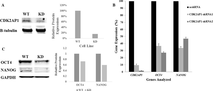
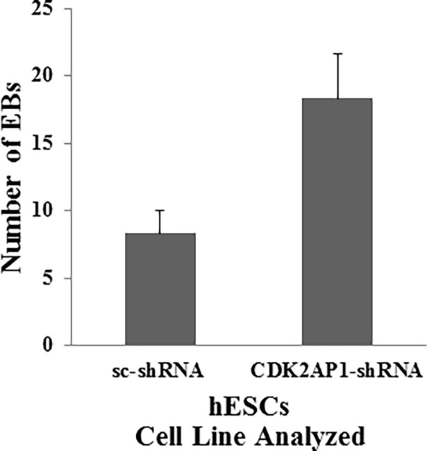
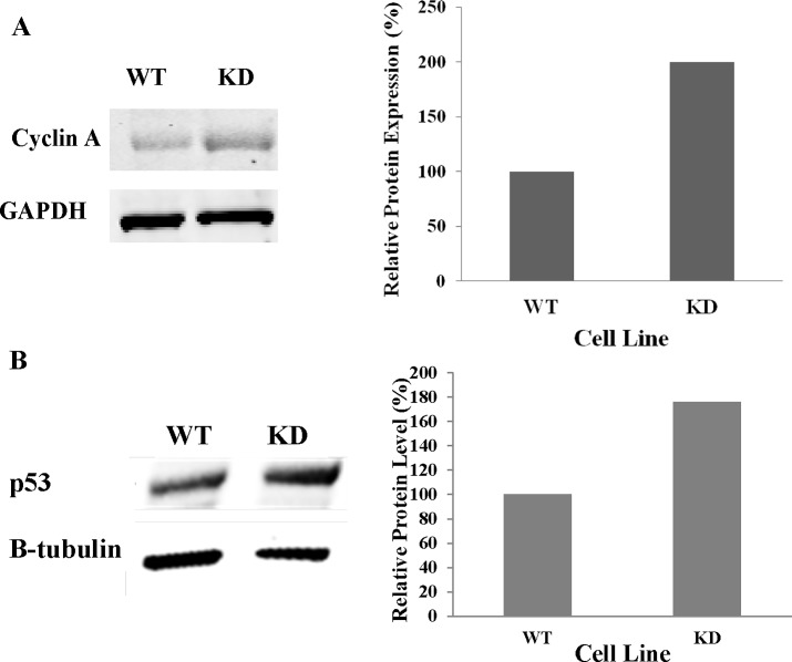
Recent studies have suggested a role for the Cyclin Dependent Kinase-2 Associated Protein 1 (CDK2AP1) in stem cell differentiation and self-renewal. In studies with mouse embryonic stem cells (mESCs) derived from generated mice embryos with targeted deletion of the Cdk2ap1 gene, CDK2AP1 was shown to be required for epigenetic silencing of Oct4 during differentiation, with deletion resulting in persistent self-renewal and reduced differentiation potential. Differentiation capacity was restored in these cells following the introduction of a non-phosphorylatible form of the retinoblastoma protein (pRb) or exogenous Cdk2ap1. In this study, we investigated the role of CDK2AP1 in human embryonic stem cells (hESCs). Using a shRNA to reduce its expression in hESCs, we found that CDK2AP1 knockdown resulted in a significant reduction in the expression of the pluripotency genes, OCT4 and NANOG. We also found that CDK2AP1 knockdown increased the number of embryoid bodies (EBs) formed when differentiation was induced. In addition, the generated EBs had significantly higher expression of markers of all three germ layers, indicating that CDK2AP1 knockdown enhanced differentiation. CDK2AP1 knockdown also resulted in reduced proliferation and reduced the percentage of cells in the S phase and increased cells in the G2/M phase of the cell cycle. Further investigation revealed that a higher level of p53 protein was present in the CDK2AP1 knockdown hESCs. In hESCs in which p53 and CDK2AP1 were simultaneously downregulated, OCT4 and NANOG expression was not affected and percentage of cells in the S phase of the cell cycle was not reduced. Taken together, our results indicate that the knockdown of CDK2AP1 in hESCs results in increased p53 and enhances differentiation and favors it over a self-renewal fate.


Conflict of interest statement

Competing Interests: The authors have declared that no competing interests exist.

Figures

Fig 1
Fig 1. Knockdown of CDK2AP1 in hESCs reduces OCT4 and NANOG expression.
A. Whole cell lysates were used to examine levels of CDK2AP1 protein in WA09 hESCs transduced with CDK2AP1 specific shRNA1 (right lane) or a control (left). Cells that were transduced with CDK2AP1 specific shRNA1 had lower CDK2AP1 protein levels. Densitometric analysis of the protein levels of the Western blot is presented in the right panel. B. WA09 hESCs that were transduced with CDK2AP1-shRNA1, CDK2AP1-shRNA2 or a scrambled shRNA (scshRNA) were examined for CDK2AP1 knockdown and for OCT4 and NANOG expression. Knockdown of CDK2AP1 using CDK2AP1-shRNA1 and CDK2AP1-shRNA2 resulted in reduction in OCT4 and NANOG expression when compared with hESCs that were transduced with the scshRNA(p-value < 0.05). Results are presented together with standard deviation from experiments conducted in triplicate. C. Whole cell lysates from WA09 hESCs transduced with CDK2AP1-shRNA1 (right lane) or scshRNA(left) were examined by Western Analysis and demonstrated reduction in OCT4 and NANOG levels. Densitometric analysis of the protein levels of the Western blot is presented in the right panel.
Fig 2
Fig 2. Knockdown of CDK2AP1 enhances differentiation.
Knockdown of CDK2AP1 in hESC increased the number of generated EBs (p < 0.05). Results are presented together with standard deviation from experiments conducted in triplicate.
Fig 3
Fig 3. Knockdown of CDK2AP1 in hESC reduces proliferation and increases cells in the G2/M and decreases cells in the S phase of the cell cycle.
A. Fifty thousand WA09 hESCs that were transduced with a scshRNA (WT) or CDK2AP1 shRNA1 (KD) were seeded per well in a 12-well plate. Cells were harvested and counted in triplicates at 2 and 4 days post seeding. (*- p-value < 0.05). Comparisons were made between WT and KD cells at respective time points). Results are presented together with standard deviation from experiments conducted in triplicate. B. CDK2AP1 wild type and knockdown WA09 hESCs were harvested and equal numbers were stained with propidium iodide (PI) and analyzed using an Accuri C6 flow cytometer. Results are presented together with standard deviation from experiments conducted in triplicate. (* = p < 0.05).
Fig 4
Fig 4. Knockdown of CDK2AP1 in hESCs increases Cyclin A and p53 protein levels.
A. Western blot (left) shows an increase in Cyclin A levels in the CDK2AP1 knockdown hESCs when compared to wild type cells. Densitometric analysis of the protein levels of the Western blot is presented in the right panel. B. Whole cell lysates of wild type and CDK2AP1 knockdown WA09 hESCs were examined for p53 protein levels. We found that knockdown of CDK2AP1 increased p53 protein levels as shown by the western blot (left). Densitometric analysis of the protein level is presented in the right panel.
Fig 5
Fig 5. Simultaneous knockdown of CDK2AP1 and p53 prevents the reduction in OCT4 and NANOG levels and the accumulation in the G2/M phase of the cell cycle.
A. BG01v hESCs were transduced with scrambled shRNA (sc-shRNA), CDK2AP1 shRNA or co-transduced with CDK2AP1 and p53 shRNAs. The gene expression of CDK2AP1, OCT4, NANOG and p53 was analyzed in those cells by qPCR. We found that down regulating p53 with CDK2AP1 prevented the CDK2AP1-knockdown induced reduction in OCT4 and NANOG levels (p < 0.05). Results are presented together with standard deviation from experiments conducted in triplicate. B. When p53 is simultaneously downregulated with CDK2AP1, there was no reduction in cells in the S phase and no increase in cells in the G2/M phase of the cell cycle. Around 6% of the analyzed cells were in the sub G0/G1 phase of the cell cycle. Results are presented together with standard deviation from experiments conducted in triplicate. Cell cycle data of scshRNA and CDK2AP1-shRNA transduced hESCs from Fig 3B were included in this figure to facilitate comparison.

References

    1. Ramalho-Santos M, Yoon S, Matsuzaki Y, Mulligan RC, Melton DA. "Stemness": transcriptional profiling of embryonic and adult stem cells. Science. 2002;298(5593):597–600. doi: 10.1126/science.1072530 - DOI - PubMed
    1. Rao RR, Calhoun JD, Qin X, Rekaya R, Clark JK, Stice SL. Comparative transcriptional profiling of two human embryonic stem cell lines. Biotechnol Bioeng. 2004;88(3):273–86. doi: 10.1002/bit.20245 - DOI - PubMed
    1. Rao RR, Stice SL. Gene expression profiling of embryonic stem cells leads to greater understanding of pluripotency and early developmental events. Biol Reprod. 2004;71(6):1772–8. doi: 10.1095/biolreprod.104.030395 - DOI - PubMed
    1. Sharov AA, Piao Y, Matoba R, Dudekula DB, Qian Y, VanBuren V, et al. Transcriptome analysis of mouse stem cells and early embryos. PLoS Biol. 2003;1(3):E74 doi: 10.1371/journal.pbio.0000074 - DOI - PMC - PubMed
    1. Kim Y, McBride J, Kimlin L, Pae EK, Deshpande A, Wong DT. Targeted inactivation of p12, CDK2 associating protein 1, leads to early embryonic lethality. PLoS One. 2009;4(2):e4518 doi: 10.1371/journal.pone.0004518 - DOI - PMC - PubMed

Publication types

MeSH terms