. 2018 May 6;10(5):244.
doi: 10.3390/v10050244.

An Ointment Consisting of the Phage Lysin LysGH15 and Apigenin for Decolonization of Methicillin-Resistant Staphylococcus aureus from Skin Wounds

Affiliations

An Ointment Consisting of the Phage Lysin LysGH15 and Apigenin for Decolonization of Methicillin-Resistant Staphylococcus aureus from Skin Wounds

Mengjun Cheng et al. Viruses. .

Abstract

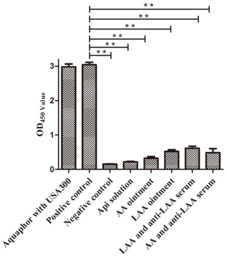
Staphylococcus aureus (S. aureus) is a common and dangerous pathogen that causes various infectious diseases. Skin damage, such as burn wounds, are at high risk of Staphylococcus aureus colonization and infection, which increases morbidity and mortality. The phage lysin LysGH15 exhibits highly efficient lytic activity against methicillin-resistant S. aureus (MRSA) and methicillin-susceptible S. aureus (MSSA) strains. Apigenin (api) significantly decreases haemolysis of rabbit erythrocytes caused by S. aureus and shows anti-inflammatory function. LysGH15 and api were added to Aquaphor to form an LysGH15-api-Aquaphor (LAA) ointment. The LAA ointment simultaneously exhibited bactericidal activity against S. aureus and inhibited haemolysis. In an LAA-treated mouse model of an MRSA-infected skin wound, the mean bacterial colony count decreased to approximately 10² CFU/mg at 18 h after treatment (and the bacteria became undetectable at 96 h), whereas the mean count in untreated mice was approximately 10⁵ CFU/mg of tissue. The LAA ointment also reduced the levels of pro-inflammatory cytokines (TNF-α, IL-1β, and IFN-γ) and accelerated wound healing in the mouse model. These data demonstrate the potential efficacy of a combination of LysGH15 and api for use as a topical antimicrobial agent against S. aureus.

Keywords: apigenin; methicillin-resistant Staphylococcus aureus; ointment; phage lysin; skin infection.


Conflict of interest statement

The authors declare no conflict of interest.

Figures

Figure 1
Figure 1
In vitro efficiency of LAA ointment. (A) Plate lytic assay. The antibacterial activity of the different ointments against the MRSA strain USA300 was tested using plate assays. The antibacterial rings were measured for the following formulations: the (a) LysGH15 solution (120 μg per 100 μL PBS), (b) Aquaphor containing LysGH15 (600 μg per 1 g ointment), (c) Aquaphor containing both LysGH15 (600 μg per 1 g ointment) and api (5000 μg per 1 g ointment), (d), api solution (100 μg per 100 μL PBS), (e) Aquaphor containing api (5000 μg per 1 g ointment), (f) pure Aquaphor (0.1 g was dripped on BHI plates), and (g) mupirocin (0.05 mg) dripped on the plates. (B) Time kill assay. The following formulations were added to cultures of the MRSA strain USA300: LAA, LA, AA, api, LysGH15, mupirocin, and Aquaphor. CFU numbers were counted at different time points as indicated (n = 3). The experimental data were analysed by using a one-way analysis of variance. * p < 0.05 compared with the buffer control. The data are representative of three experiments.
Figure 2
Figure 2
SEM observations of the highly efficient lytic activity of LAA. USA300 was treated with the following formulations: (a) Aquaphor, (b) api solution, (c) LysGH15 solution, (d) LA ointment, and (e) LAA ointment. The images were obtained by SEM. The bars indicate 1 μm.
Figure 3
Figure 3
Haemolytic assays. The OD450 of the different supernatants was measured. The negative control was PBS-treated erythrocyte suspensions. The experimental data were analysed by using a one-way analysis of variance. ** p < 0.01 compared with the positive control. The data are representative of three experiments.
Figure 4
Figure 4
LAA promotes skin wound healing. The shaved and tape-stripped mice were infected with USA300 (1 × 10⁷ CFU/mouse). At 24 h after colonization, the mice were topically treated with different formulations. (A) Photographs of wound sites treated with different formulations: the (a) PBS-treated group, (b) Aquaphor-treated group, (c) AA ointment-treated group, (d) LA ointment-treated group, (e) LAA ointment-treated group, and (f) mupirocin-treated group. After treatment, the wound sites were photographed at the 1, 3, 5, 7 and 9 days. (B) Wound healing rates. Percentage wound closure in the groups treated with different formulations: PBS-treated, Aquaphor-treated, AA ointment-treated, LA ointment-treated, LAA ointment-treated, and mupirocin-treated. The experimental data were analysed by using a one-way analysis of variance. * p < 0.05 compared with the PBS-treated control. (C) Levels of mRNA relative to GAPDH for the AAM-associated genes arginase I and YM1 at 5 d after injury. Alternatively activated macrophages (AAMs) were induced in the skin wounds of LAA-treated mice at 5 d after treatment (n = 6 mice per group per experiment). The experimental data was analysed by using a one-way analysis of variance. * p < 0.05 compared with the PBS-treated control.
Figure 5
Figure 5
Histological re-epithelialization of skin wounds. The mice were monitored and treated. (a) normal mouse skin, (b) PBS-treated mouse skin, (c) Aquaphor-treated mouse skin, (d) AA-treated mouse skin, (e) LA-treated mouse skin, (f) LAA-treated mouse skin, and (g) mupirocin-treated mouse skin. n = 6 mice per group per experiment. The skin wounds were stained with haematoxylin and eosin (100×). The arrows indicate the hair follicles (I) and the cortical layer (II) of the skin wounds. Scale bars = 500 μm.
Figure 6
Figure 6
In vivo decolonization activity of LAA in tape-stripped mice infected with MRSA. The tape-stripped mice were infected with MRSA USA300 (1 × 10⁷ CFU/mouse). At 24 h after colonization, the mice were treated with the topical treatments. The bacterial load in the wounded skin at 18 h after treatment was assessed; n = 6 mice per group per experiment. The experimental data was analysed by using a one-way analysis of variance. Compared with the PBS-treated group: ** p < 0.01. The median value for each group is represented as a horizontal bar, and each sphere represents one mouse. The dotted line indicated the low limit of detection line.
Figure 7
Figure 7
LAA reduced levels of pro-inflammatory cytokines. The levels of TNF-α, IFN-γ, and IL-1β in the excised wound area were determined at 18 h after topical treatment (n = 6 mice per group per experiment). The experimental data was analysed by using a one-way analysis of variance. Compared with the control group: ## p < 0.01. Compared with the PBS-treated group: * p < 0.05. Compared with the control group and PBS-treated group: *# p < 0.05.
Figure 8
Figure 8
Anti-LAA serum did not neutralize the lytic activity of LAA in vitro. Serum samples from the mice that were exposed to one dose per day of topical LAA for 9 d and the control mice were collected at 10 d after treatment. (A) The titres were measured using ELISA. Serum from LAA-treated mice was employed as the primary antibody, and HRP-labelled anti-mouse IgG antibody was employed as the secondary antibody. Western blotting assays of the LysGH15 antibody were carried out (n = 6 mice per group per experiment). (B) The influence of anti-LAA serum on the lytic activity of LAA. CFUs were counted at different time points, as indicated. Circle: Aquaphor. Box: LAA. Inverted triangle: anti-LAA serum with LAA. n = 3 per group per experiment. The experimental data was analysed by using a one-way analysis of variance. * p < 0.05 compared with PBS-treated mice. The data are representative of three experiments.

References

    1. Peacock J.J., Marsik F.J., Wenzel R.P. Methicillin-resistant Staphylococcus aureus: Introduction and spread within a hospital. Ann. Intern. Med. 1980;93:526–532. doi: 10.7326/0003-4819-93-4-526. - DOI - PubMed
    1. Tong S.Y., Davis J.S., Eichenberger E., Holland T.L., Fowler V.J. Staphylococcus aureus infections: Epidemiology, pathophysiology, clinical manifestations, and management. Clin. Microbiol. Rev. 2015;28:603–661. doi: 10.1128/CMR.00134-14. - DOI - PMC - PubMed
    1. Iruloh C.G., Bonner S., Ma K. Methicillin-sensitive Staphylococcus aureus chorioamnionitis and foetal death after mechanical induction of labour: A case report. J. Obstet. Gynaecol. 2017;38:1–2. doi: 10.1080/01443615.2017.1343809. - DOI - PubMed
    1. Sasson G., Bai A.D., Showler A., Burry L., Steinberg M., Ricciuto D.R., Fernandes T., Chiu A., Raybardhan S., Science M., et al. Staphylococcus aureus bacteremia in immunosuppressed patients: A multicenter, retrospective cohort study. Eur. J. Clin. Microbiol. Infect. Dis. 2017;36:1231–1241. doi: 10.1007/s10096-017-2914-y. - DOI - PubMed
    1. Fischetti V.A. Novel Method to Control Pathogenic Bacteria on Human Mucous Membranes. Ann. N. Y. Acad. Sci. 2003;987:207–214. doi: 10.1111/j.1749-6632.2003.tb06050.x. - DOI - PubMed

Publication types

MeSH terms