. 2018 May 22;115(21):5468-5473.
doi: 10.1073/pnas.1709211115. Epub 2018 May 7.

Defective phagosome motility and degradation in cell nonautonomous RPE pathogenesis of a dominant macular degeneration

Affiliations

Defective phagosome motility and degradation in cell nonautonomous RPE pathogenesis of a dominant macular degeneration

Julian Esteve-Rudd et al. Proc Natl Acad Sci U S A. .

Abstract

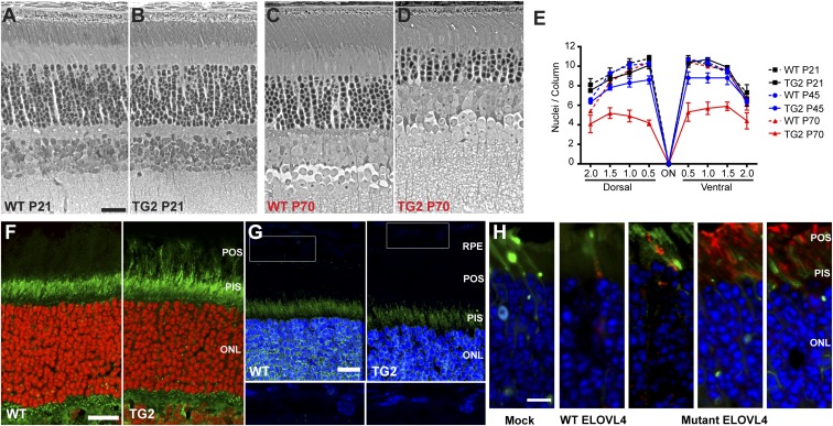
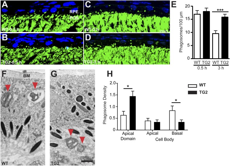
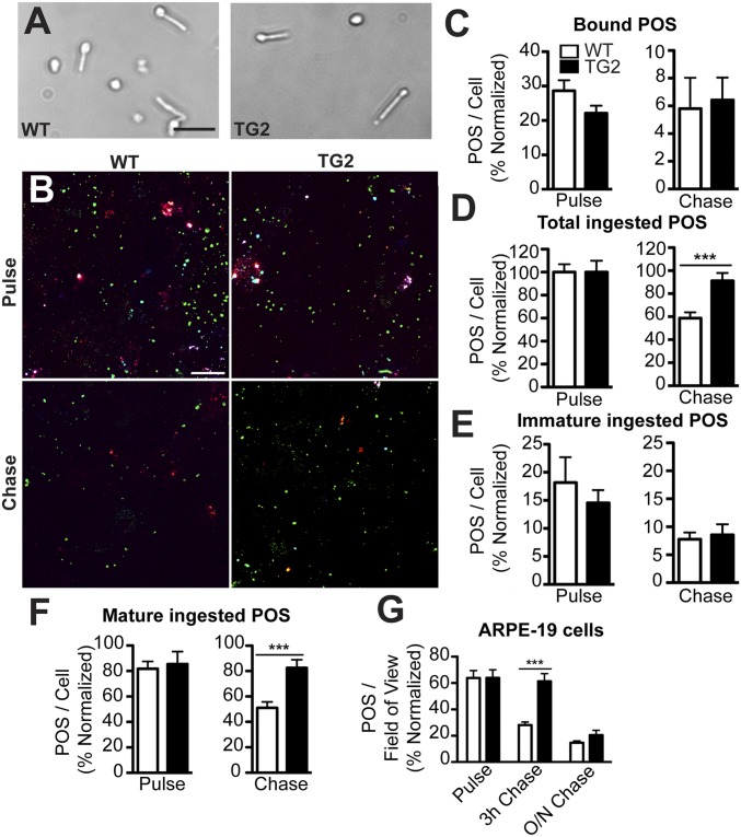
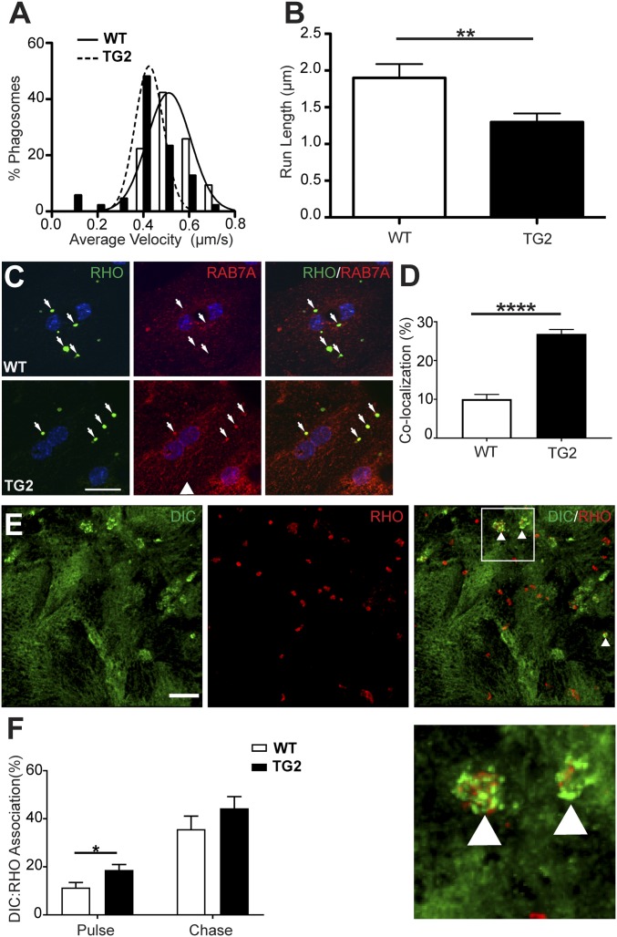
Stargardt macular dystrophy 3 (STGD3) is caused by dominant mutations in the ELOVL4 gene. Like other macular degenerations, pathogenesis within the retinal pigment epithelium (RPE) appears to contribute to the loss of photoreceptors from the central retina. However, the RPE does not express ELOVL4, suggesting photoreceptor cell loss in STGD3 occurs through two cell nonautonomous events: mutant photoreceptors first affect RPE cell pathogenesis, and then, second, RPE dysfunction leads to photoreceptor cell death. Here, we have investigated how the RPE pathology occurs, using a STGD3 mouse model in which mutant human ELOVL4 is expressed in the photoreceptors. We found that the mutant protein was aberrantly localized to the photoreceptor outer segment (POS), and that resulting POS phagosomes were degraded more slowly in the RPE. In cell culture, the mutant POSs are ingested by primary RPE cells normally, but the phagosomes are processed inefficiently, even by wild-type RPE. The mutant phagosomes excessively sequester RAB7A and dynein, and have impaired motility. We propose that the abnormal presence of ELOVL4 protein in POSs results in phagosomes that are defective in recruiting appropriate motor protein linkers, thus contributing to slower degradation because their altered motility results in slower basal migration and fewer productive encounters with endolysosomes. In the transgenic mouse retinas, the RPE accumulated abnormal-looking phagosomes and oxidative stress adducts; these pathological changes were followed by pathology in the neural retina. Our results indicate inefficient phagosome degradation as a key component of the first cell nonautonomous event underlying retinal degeneration due to mutant ELOVL4.

Keywords: ELOVL4; Stargardt; phagocytosis; photoreceptor.


Conflict of interest statement

The authors declare no conflict of interest.

Figures

Fig. 1.
Fig. 1.
Retinal degeneration and localization of ELOVL4. (AD) Phase-contrast images of semithin sections from P21 or P70 WT (A and C) and TG2 (B and D) retinas. Mutant TG2 retinas were similar in overall morphology to WT retinas at P21, but the photoreceptor cells have undergone markedly significant degeneration by P70, as can be observed by the loss of photoreceptor nuclei in the outer nuclear layer (ONL). (E) Graph of the number of photoreceptor nuclei per column in the ONL, quantified at 0.5-mm intervals from the optic nerve (ON). Some photoreceptor cell loss in TG2 retinas was observed at P45; by P70 it was more severe. (F and G) Retinal sections from P21 WT and TG2 mice immunolabeled with antibodies against the N-terminal (F) or C-terminal (G) region of ELOVL4 (green). The N-terminal antibody recognizes both WT and mutant ELOVL4, but the C-terminal antibody recognizes only WT ELOVL4. Photoreceptor outer segments (POSs) are labeled in the TG2 retinal section by the former (F), but not the latter (G). Nuclei were counterstained with DAPI (shown as red or blue). Lower panels (G) represent higher magnification of rectangles in Upper panels, with the brightness of the blue channel increased to make weaker DAPI staining of RPE cells visible. (H) Retinal sections from mice whose photoreceptors were electroporated with Dendra2 (green) and FLAG-tagged WT or mutant human ELOVL4 plasmids. The sections were labeled with a FLAG antibody (red). Mock retinas were electroporated with Dendra2 only. The WT FLAG-ELOVL4 protein is localized primarily to the photoreceptor inner segment (PIS). The three panels to the Right are examples from different experiments with the mutant FLAG-ELOVL4; protein was observed mostly in the POS, with some presence in the PIS (mutant ELOVL4 still contains the N-terminal ER localization signal, but it has lost the C-terminal ER retention signal). (Scale bars: 10 μm in A, F, and H, and 15 μm in G.)
Fig. 2.
Fig. 2.
Degradation of POS phagosomes in WT and mutant mouse RPE in vivo. (AD) Immunofluorescence images of RHO labeling (green) in WT (A and C) and TG2 (B and D) retinal sections from P21 mice killed 0.5 h (A and B) and 3 h (C and D) post light onset. POS phagosomes can be seen as green particles in the RPE layer. The nuclei are counterstained with DAPI (blue). (E) Quantification of POS phagosomes per 100 μm of the RPE layer in WT and TG2 retinas showed no significant difference at 0.5 h post light onset, but significantly more POS phagosomes remained in TG2 RPE at 3 h post light onset, indicating impaired phagosome degradation. (F and G) ImmunoEMs of WT (F) and TG2 (G) retinas from P28 mice showing POS phagosomes (immunogold labeled with RHO antibodies, and indicated by red arrowheads) inside the RPE at 1.5 h post light onset. (H) Quantification of phagosome density (mean number of phagosomes per 55 μm2 of sectioned RPE) in different regions of the RPE. Bar graph shows data from the apical and cell body domains of the RPE; these domains border each other at the level of the junctional complexes. The cell body domain was divided further into apical and basal halves. The data show that more TG2 than WT POS phagosomes were in the apical domain, while more WT than TG2 phagosomes were in the basal half of the cell body. BM, Bruch’s membrane. (Scale bars in C and G: 10 μm and 1 μm, respectively.) Error bars in E and H represent ±SEM. *P < 0.05; ***P < 0.001.
Fig. 3.
Fig. 3.
Impaired degradation of mutant ELOVL4 POSs by WT RPE cultures. (A) Brightfield images of WT and TG2 POSs isolated from respective retinas. (B) RHO labeling of WT primary mouse RPE cells, fed either WT or TG2 POSs, immediately following the pulse or following a chase period. The labeling was performed before and after cell permeabilization to distinguish between surface-bound POSs (white or red) and internalized phagosomes. Additionally, the immature phagosomes (cyan) were identified by labeling with a RHO mAb that binds the C terminus (1D4) and RHO pAb01, while the mature phagosomes (green) were identified by labeling with RHO pAb01 only. (CF) Quantification of the POSs. POSs fed to primary mouse RPE and labeled with RHO pAb01, showed that there was no significant difference between the number of bound POSs after the pulse and chase (C), but there were more total ingested TG2 POSs following the chase compared with WT POSs (D). When the total POSs were further defined as immature (labeled with mAb1D4) vs. mature (labeled with pAb01 but not mAb1D4), the number of immature ingested POSs was not found to be statistically significant between WT and TG2 POSs (E), whereas the cells fed TG2 POSs had more mature ingested POSs following the chase (F). (G) The impaired POS degradation phenotype was also observed in human ARPE-19 cells, which after a 3-h chase period, had more (bound + ingested) TG2 POSs than ARPE-19 cells fed WT POSs. O/N, overnight. (Scale bars in A and B: 20 μm and 10 μm, respectively.) Error bars in CG represent ±SEM. ***P < 0.001.
Fig. 4.
Fig. 4.
Impaired motility of TG2 phagosomes in WT primary mouse RPE cells. (A and B) Live-cell analysis of the tracks of phagosomes (85 per condition), from either WT or TG2 POSs, showed that TG2 phagosomes had a lower mean velocity (A) and shorter mean run length (B) than their WT counterparts. (C) Immunolabeling of RHO (green) and the endosomal marker RAB7A (red) in WT primary mouse RPE cells, fed either WT or TG2 POSs. (D) Quantification of the number of RHO-positive phagosomes colocalized with RAB7A showed that more phagosomes, derived from TG2 POSs relative to WT POSs, had RAB7A associated with them after a 1-h chase period. (E) Immunofluorescence of cytoplasmic dynein intermediate chain (DIC, green) and RHO (red) in WT primary mouse RPE cells fed POSs after a 1-h chase. White arrowheads in the merged panel indicate phagosomes associated with DIC immunolabeling, shown at higher magnification Below. (F) Quantification of the proportion of WT or TG2 POS phagosomes in WT primary mouse RPE cells that were associated with DIC immunolabel, after a 20-min pulse and a 1-h chase. (Scale bars in C and E: 20 μm and 5 μm, respectively.) Error bars in B, D, and F represent ±SEM. *P < 0.05; **P < 0.01; ****P < 0.0001.

References

    1. Zhang K, et al. A 5-bp deletion in ELOVL4 is associated with two related forms of autosomal dominant macular dystrophy. Nat Genet. 2001;27:89–93. - PubMed
    1. Edwards AO, Donoso LA, Ritter R., 3rd A novel gene for autosomal dominant Stargardt-like macular dystrophy with homology to the SUR4 protein family. Invest Ophthalmol Vis Sci. 2001;42:2652–2663. - PubMed
    1. Agbaga MP, et al. Role of Stargardt-3 macular dystrophy protein (ELOVL4) in the biosynthesis of very long chain fatty acids. Proc Natl Acad Sci USA. 2008;105:12843–12848. - PMC - PubMed
    1. Bernstein PS, et al. Diverse macular dystrophy phenotype caused by a novel complex mutation in the ELOVL4 gene. Invest Ophthalmol Vis Sci. 2001;42:3331–3336. - PubMed
    1. Maugeri A, et al. A novel mutation in the ELOVL4 gene causes autosomal dominant Stargardt-like macular dystrophy. Invest Ophthalmol Vis Sci. 2004;45:4263–4267. - PubMed

Publication types

MeSH terms