

Addition of ultrasound to mammography in the case of dense breast tissue: systematic review and meta-analysis
- PMID: 29736009
- PMCID: PMC6008336
- DOI: 10.1038/s41416-018-0080-3
Addition of ultrasound to mammography in the case of dense breast tissue: systematic review and meta-analysis
Abstract
Background: Mammography is less effective in detecting cancer in dense than in fatty breasts.
Methods: We undertook a systematic search in PubMed to identify studies on women with dense breasts who underwent screening with mammography supplemented with ultrasound. A meta-analysis was undertaken on the proportion of cancers detected only by ultrasound, out of all screen-detected cancers, and the proportion of women with negative mammography who were referred for assessment following ultrasound screening.
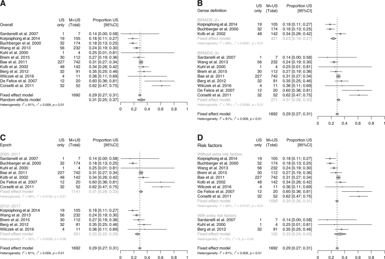
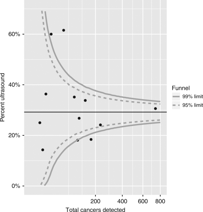
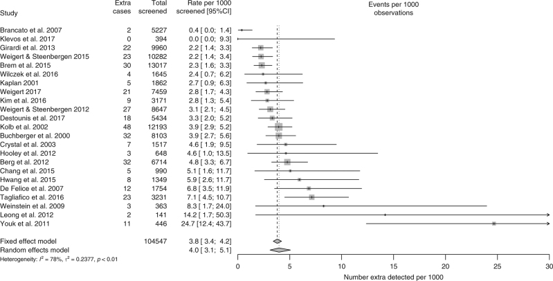
Results: Twenty-nine studies satisfied our inclusion criteria. The proportion of total cancers detected only by ultrasound was 0.29 (95% CI: 0.27-0.31), consistent with an approximately 40% increase in the detection of cancers compared to mammography. In the studied populations, this translated into an additional 3.8 (95% CI: 3.4-4.2) screen-detected cases per 1000 mammography-negative women. About 13% (32/248) of cancers were in situ from 17 studies with information on this subgroup. Ultrasound approximately doubled the referral for assessment in three studies with these data.
Conclusions: Studies have consistently shown an increased detection of breast cancer by supplementary ultrasound screening. An inclusion of supplementary ultrasound into routine screening will need to consider the availability of ultrasound and diagnostic assessment capacities.
Conflict of interest statement
The authors declare no competing interests.
Figures

Comment in
-
Comment on 'Addition of ultrasound to mammography in the case of dense breast tissue: systematic review and meta-analysis'.Br J Cancer. 2018 Nov;119(11):1443. doi: 10.1038/s41416-018-0237-0. Epub 2018 Nov 9. Br J Cancer. 2018. PMID: 30410057 Free PMC article. No abstract available.
-
Reply to 'Comment on 'Addition of ultrasound to mammography in the case of dense breast tissue: systematic review and meta-analysis".Br J Cancer. 2018 Nov;119(11):1444. doi: 10.1038/s41416-018-0247-y. Epub 2018 Nov 9. Br J Cancer. 2018. PMID: 30410058 Free PMC article. No abstract available.
References
-
- Tabar L, et al. Update of the Swedish two-county program of mammographic screening for breast cancer. Radiol. Clin. North Am. 1992;30:187–210. - PubMed
-
- International Agency for research on Cancer. Breast Cancer Screening (IARC Handbooks of Cancer Prevention), Vol. 15 (IARC, Lyon, France, 2016). .
-
- Perry, N. B. et al. (eds). European Guidelines for Quality Assurance in Breast Cancer Screening and Diagnosis, 4th edn (Office for Official Publications of the European Communities, Luxembourg, 2006).
Publication types
MeSH terms
Grants and funding
LinkOut - more resources
Full Text Sources
Other Literature Sources
Medical
