. 2018 Mar 15:2018:9787239.
doi: 10.1155/2018/9787239. eCollection 2018.

Decreased IL-33 Production Contributes to Trophoblast Cell Dysfunction in Pregnancies with Preeclampsia

Affiliations

Decreased IL-33 Production Contributes to Trophoblast Cell Dysfunction in Pregnancies with Preeclampsia

Hong Chen et al. Mediators Inflamm. .

Abstract

Preeclampsia (PE) is a life-threatening pregnancy complication which is related to aggradation of risk regarding fetal and maternal morbidity and mortality. Dysregulation of systemic inflammatory response and dysfunction of trophoblast cells have been proposed to be involved in the development and progression of PE. Some studies have demonstrated that interleukin-33 (IL-33) is an immunomodulatory cytokine that is associated with the immune regulation of tumor cells. However, little is known whether IL-33 and its receptor ST2/IL-1 R4 could regulate trophoblast cells, which are associated with the pathogenesis of PE. In this study, our target is to explore the impact of IL-33 on trophoblast cells and elucidate its underlying pathophysiological mechanisms. Placental tissues from the severe PE group (n = 11) and the normotensive pregnant women's group (n = 11) were collected for the protein expression and distribution of IL-33 along with its receptor ST2/IL-1 R4 via Western blot analysis and immunohistochemistry, respectively. We discovered that the level of IL-33 was decreased in placental tissues of pregnant women with PE, while no distinction was observed in the expression of ST2/IL-1 R4. These results were further verified in villous explants which were treated with sodium nitroprusside with different concentrations, to simulate the pathological environment of PE. To investigate IL-33 effects on trophoblast cells separately, IL-33 shRNA was introduced into HTR8/SVneo cells and villi. IL-33 shRNA weakened the proliferation, migration, and invasion capacity of HTR8/SVneo cells. The migration distance of villous explants was also markedly decreased. The reduced invasion of trophoblast cells is a result of IL-33 knockdown which could be related to the decline of MMP2/9 activity and the increased utterance of TIMP1/2. Overall, our findings demonstrated that the reduction of IL-33 production was connected with the reduced functional capability of trophoblast cells, thus inducing placental insufficiency that has been linked to the development of PE.


Figures
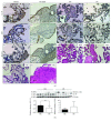
Figure 1
Figure 1
Expression and distribution of IL-33 and ST2/IL-1 R4 in placental tissues. (a) IL-33 was localized in syncytiotrophoblast (STB) cells, cytotrophoblast (CTB) cells, trophoblast columns (TC) of placental villi (A–C), and some extravillous trophoblasts (EVT) in maternal decidual cells (E–G). D, H, K, and N were the negative controls (NEG). CK7 is a marker for STB and TC. HLA-G is a marker for EVT. The IL-33 in women with normal (I, J) or preeclamptic pregnancies (L, M) was localized in trophoblast cells (200x; scale bar, 200 μm). (b) The expression of IL-33 is decreased remarkably in women complicated with preeclampsia, while the expression of ST2/IL-1 R4 in placenta shows no difference between these two groups (t-test, ∗∗ P = 0.0057).
Figure 2
Figure 2
Western blot demonstrating the level of IL-33 in villous explants treated with SNP at different concentrations and immunofluorescence images demonstrating the expression of ST2/IL-1 R4 in these groups. (a) The level of IL-33 was high in low concentrations of SNP and then decreased continuously with increasing concentrations of SNP. (b, c) Fluorescence specific to ST2/IL-1 R4 is red, and the nuclei were stained by DAPI (blue) (200x; scale bar, 200 μm). ST2/IL-1 R4, the receptor of IL-33, showed no significant differences in these groups (t-test, P < 0.05).
Figure 3
Figure 3
Immunofluorescence images, Western blot, and qRT-PCR were used to evaluate the transfection efficiency of shRNA targeting IL-33 in HTR8/SVneo cells. (a) Fluorescence specific to IL-33 is red, and the nuclei were stained by DAPI (blue) (400x; scale bar, 100 μm). (b, c) The transfection efficiency of IL-33 knockdown by shRNA is shown by Western blot and qRT-PCR (t-test, ∗∗ P < 0.01, ∗∗∗ P < 0.001).
Figure 4
Figure 4
Treatment with IL-33 shRNA weakened the proliferation, invasion, and migration of HTR8/SVneo cells. (a, b) IL-33 knockdown significantly decreased the area of the wound in the wound healing assay (40x; scale bar, 50 μm). (c) IL-33 knockdown significantly weakened the proliferation of cells. (d–f) The migration and invasion potency of HTR8/SVneo cells was decreased in the IL-33 knockdown group compared with scrambled shRNA group (200x; scale bar, 200 μm) (t-test, ∗∗ P < 0.01, ∗∗∗ P < 0.001).
Figure 5
Figure 5
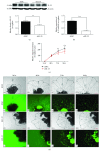
Knockdown of IL-33 decreased the outgrowth of the human villous explants. (a, b) Western blot and qRT-PCR were used to evaluate the transfection efficiency of shRNA targeting IL-33 in villous explants. (c, d) The outgrowth of villous explants treated with shRNA was photographed at 24 hr (Aa, Ee), 48 hr (Bb, Ff), 72 hr (Cc, Gg), and 96 hr (Dd, Hh) by a light microscope and fluorescence microscope (40x; scale bar, 50 μm). The migration distance was measured using ImageJ software (t-test, ∗∗ P < 0.01, ∗∗∗ P < 0.001).
Figure 6
Figure 6
IL-33 knockdown decreased the activity of MMP2/9 and increased the utterance of TIMP1/2. (a) Image of gelatin zymography assay (GZ) using the medium of HTR8/SVneo cells disturbed by shRNA. Western blot (WB) was used to test the utterance of TIMP1/2 in proteins from these HTR/SVneo cells. (b) Image of the gelatin zymography assay using the medium of villous explants disturbed by shRNA. Western blot was used to test the utterance of TIMP1/2 in proteins from these explants (t-test, ∗∗ P < 0.01).

References

    1. Mol B. W., Roberts C. T., Thangaratinam S., Magee L. A., de Groot C. J., Hofmeyr G. J. Pre-eclampsia. The Lancet. 2016;387(10022):999–1011. doi: 10.1016/S0140-6736(15)00070-7. - DOI - PubMed
    1. Ji L., Brkic J., Liu M., Fu G., Peng C., Wang Y. L. Placental trophoblast cell differentiation: physiological regulation and pathological relevance to preeclampsia. Molecular Aspects of Medicine. 2013;34(5):981–1023. doi: 10.1016/j.mam.2012.12.008. - DOI - PubMed
    1. Myatt L. Role of placenta in preeclampsia. Endocrine. 2002;19(1):103–111. doi: 10.1385/ENDO:19:1:103. - DOI - PubMed
    1. Knofler M., Pollheimer J. IFPA Award in Placentology lecture: molecular regulation of human trophoblast invasion. Placenta. 2012;33:S55–S62. doi: 10.1016/j.placenta.2011.09.019. - DOI - PMC - PubMed
    1. Pennington K. A., Schlitt J. M., Jackson D. L., Schulz L. C., Schust D. J. Preeclampsia: multiple approaches for a multifactorial disease. Disease Models & Mechanisms. 2012;5(1):9–18. doi: 10.1242/dmm.008516. - DOI - PMC - PubMed

MeSH terms

Substances

LinkOut - more resources