. 2018 Mar 15:2018:1289103.
doi: 10.1155/2018/1289103. eCollection 2018.

Parity-Dependent Hemosiderin and Lipofuscin Accumulation in the Reproductively Aged Mouse Ovary

Affiliations

Parity-Dependent Hemosiderin and Lipofuscin Accumulation in the Reproductively Aged Mouse Ovary

Ulises Urzua et al. Anal Cell Pathol (Amst). .

Abstract

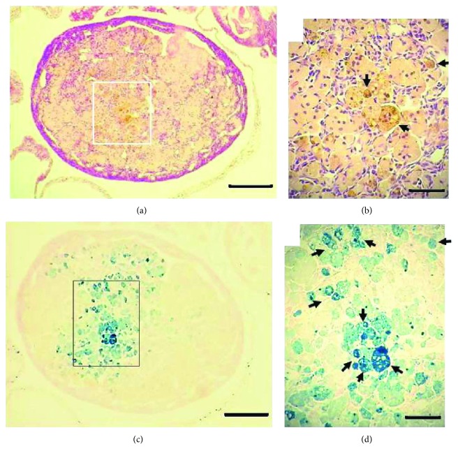
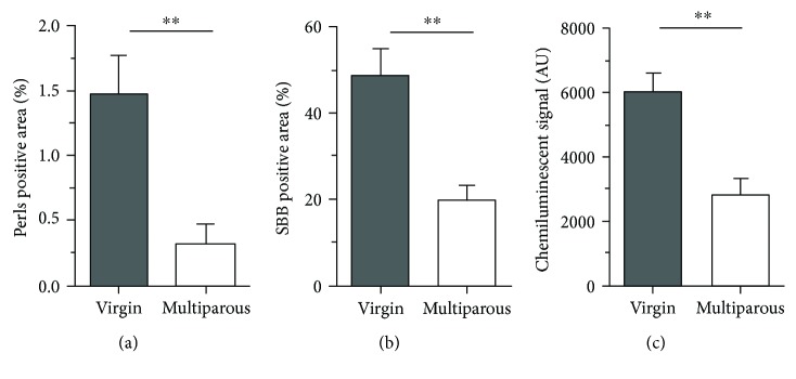
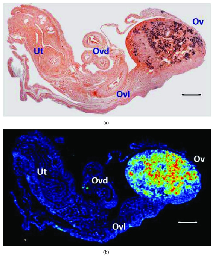
The progressive decline of the ovarian follicle pool leads to reproductive ageing. The latter is accompanied by age-related disorders, including various types of cancer. In fact, the highest rates of ovarian cancer (OC) occur at postmenopause while OC risk is significantly modulated by parity records during previous fertile life. We approached the age-parity relationship in the C57BL/6 mouse model and herein describe the presence of nonheme iron (hemosiderin) and deposits of the "age pigment" lipofuscin in reproductively aged mouse ovaries by applying conventional histochemical methods and autofluorescence. In addition, the 8-OHdG adduct was evaluated in ovarian genomic DNA. Both hemosiderin and lipofuscin were significantly higher in virgin compared to multiparous ovaries. The same pattern was observed for 8-OHdG. We conclude that nulliparity induces a long-term accumulation of iron and lipofuscin with concomitant oxidative damage to DNA in the mouse ovary. Since lipofuscin is a widely accepted senescence marker and given the recently postulated role of lipofuscin-associated iron as a source of reactive oxygen species (ROS) in senescent cells, these findings suggest a possible pathogenic mechanism by which nulliparity contributes to an increased OC risk in the postmenopausal ovary.


Figures

Figure 1
Figure 1
Hemosiderin in the aged C56BL/6 ovary. HE stain (a, b) and Perls stain (c, d) of a virgin ovary 20.6 months old. Arrows in (b) indicate brown-yellowish granules of hemosiderin. Arrows in (d) show hemosiderin laden macrophages (HLMs). Bars in (a) and (c) = 200 μm; bars in (b) and (d) = 20 μm.
Figure 2
Figure 2
Semiquantification of hemosiderin, lipofuscin, and 8-OHdG in aged ovaries. Percentages of positive signal areas of (a) Perls, n = 8 per group, and (b) SBB, n = 7 per group, relative to the total ovarian area as extracted with the ImageJ1 software. (c) 8-OHdG in isolated genomic DNA, n = 6 per group; AU = chemiluminescent arbitrary units measured with the Image Studio Digits software (for details, see Materials and Methods). ∗∗p < 0.005, nonparametric Mann–Whitney test.
Figure 3
Figure 3
Lipofuscin in the aged C56BL/6 ovary. SBB stain (a) and 543 nm green laser autofluorescence (b) of a 20.9 months old virgin ovary. Ut = uterine horn; Ovd = oviduct; Ovl = ovarian ligament; Ov = ovary. Bars in (a) and (b) = 200 μm. Image in (b) was obtained with the rainbow palette.

References

    1. Lopez-Otin C., Blasco M. A., Partridge L., Serrano M., Kroemer G. The hallmarks of aging. Cell. 2013;153(6):1194–1217. doi: 10.1016/j.cell.2013.05.039. - DOI - PMC - PubMed
    1. Broekmans F. J., Soules M. R., Fauser B. C. Ovarian aging: mechanisms and clinical consequences. Endocrine Reviews. 2009;30(5):465–493. doi: 10.1210/er.2009-0006. - DOI - PubMed
    1. Hunn J., Rodriguez G. C. Ovarian cancer: etiology, risk factors, and epidemiology. Clinical Obstetrics and Gynecology. 2012;55(1):3–23. doi: 10.1097/GRF.0b013e31824b4611. - DOI - PubMed
    1. Jeon S. Y., Hwang K. A., Choi K. C. Effect of steroid hormones, estrogen and progesterone, on epithelial mesenchymal transition in ovarian cancer development. The Journal of Steroid Biochemistry and Molecular Biology. 2016;158:1–8. doi: 10.1016/j.jsbmb.2016.02.005. - DOI - PubMed
    1. Jabara S., Christenson L. K., Wang C. Y., et al. Stromal cells of the human postmenopausal ovary display a distinctive biochemical and molecular phenotype. The Journal of Clinical Endocrinology & Metabolism. 2003;88(1):484–492. doi: 10.1210/jc.2002-021274. - DOI - PubMed

LinkOut - more resources