. 2018 Aug;391(8):833-846.
doi: 10.1007/s00210-018-1507-3. Epub 2018 May 8.

Molecular basis for the sensitivity of TRP channels to polyunsaturated fatty acids

Affiliations

Molecular basis for the sensitivity of TRP channels to polyunsaturated fatty acids

Marc Riehle et al. Naunyn Schmiedebergs Arch Pharmacol. 2018 Aug.

Abstract

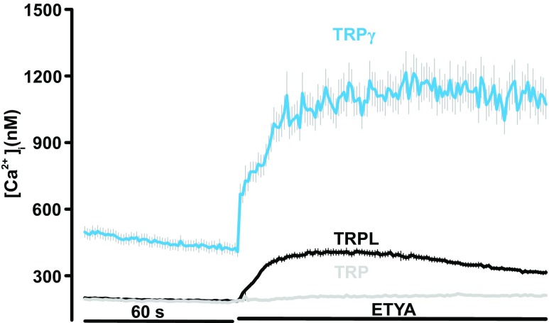
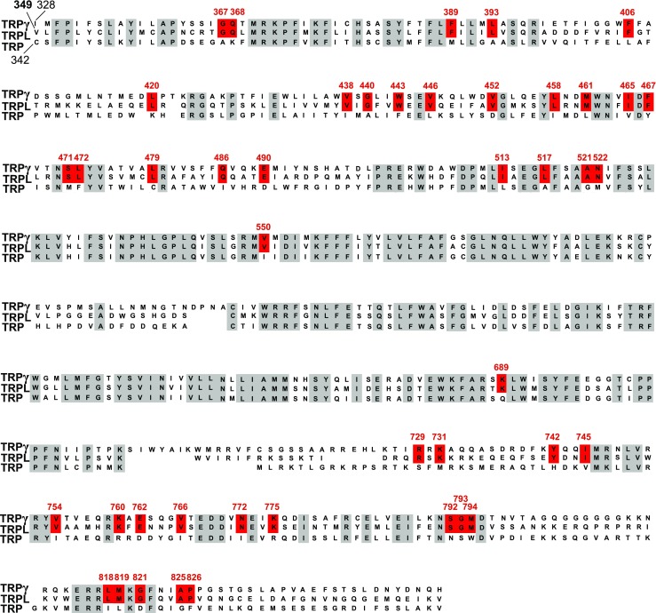
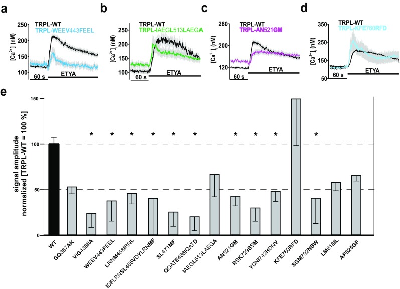
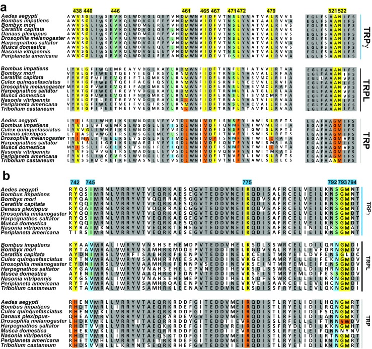
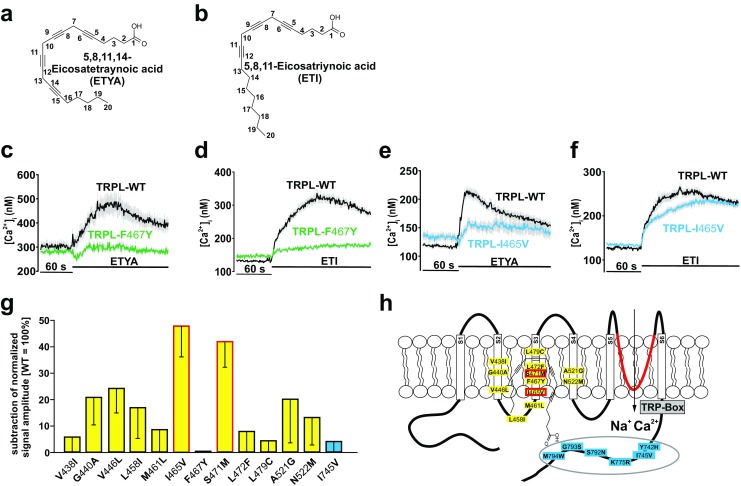
Transient receptor potential (TRP) channels represent a superfamily of unselective cation channels that are subdivided into seven subfamilies based on their sequence homology and differences in gating and functional properties. Little is known about the molecular mechanisms of TRP channel regulation, particularly of the "canonical" TRP (TRPC) subfamily and their activation by polyunsaturated fatty acids (PUFAs). Here, we analyzed the structure-function relationship of Drosophila fruit fly TRPC channels. The primary aim was to uncover the molecular basis of PUFA sensitivity of Drosophila TRP-like (TRPL) and TRPgamma channels. Amino acid (aa) sequence alignment of the three Drosophila TRPC channels revealed 50 aa residues highly conserved in PUFA-sensitive TRPL and TRPgamma channels but not in the PUFA-insensitive TRP channel. Substitution of respective aa in TRPL by corresponding aa of TRP identified 18 residues that are necessary for PUFA-mediated activation of TRPL. Most aa positions are located within a stretch comprising transmembrane domains S2-S4, whereas six aa positions have been assigned to the proximal cytosolic C-terminus. Interestingly, residues I465 and S471 are required for activation by 5,8,11,14-eicosatetraynoic acid (ETYA) but not 5,8,11-eicosatriynoic acid (ETI). As proof of concept, we generated a PUFA-sensitive TRP channel by exchanging the corresponding aa from TRPL to TRP. Our study demonstrates a specific aa pattern in the transmembrane domains S2-S4 and the proximal C-terminus essential for TRP channel activation by PUFAs.

Keywords: Ca2+ influx; Drosophila; Polyunsaturated fatty acids; TRP channels; TRPC channels.


Conflict of interest statement

The authors have no financial or other conflict of interest to disclose.

Figures

Fig. 1
Fig. 1
Functional characterization of the PUFA-induced activation of TRPgamma, TRP, and TRPL channels. Changes in intracellular calcium concentration were measured in Fura-2-loaded HEK293 expressing TRP (n = 1292), TRPL (n = 1918), and TRPgamma (n = 204) in response to application of 40 μM ETYA. Mean traces ± SEM are shown
Fig. 2
Fig. 2
Sequence alignment of the Drosophila TRPgamma, TRPL, and TRP channels. Amino acid (aa) residues over all three TRPC channels have a gray background. The aa residues preserved in TRPL and TRPgamma but not in TRPL are highlighted in red. Numbered amino acid residues correspond to the sequence of TRPL channel from Drosophila
Fig. 3
Fig. 3
Functional characterization of TRPL multiple mutants. Changes in intracellular calcium concentration over time in response to application of 40 μM ETYA in Fura-2-loaded HEK293 expressing TRPL-WT compared to TRPL multiple mutants: TRPL-WEEV443FEEL (a), TRPL-IAEGL513LAEGA (b), TRPL-AN521GM (c), and TRPL-KFE760RFD (d). Mean traces ± SEM are shown. Statistical analyses of signal amplitudes obtained during at least three independent experiments are presented as bar graphs (e). Data are means of the normalized (WT = 100%) signal amplitudes ± SEM representing the tested multiple mutants. The analysis is based on the following cell numbers: GQ367AK, n = 207; VIG438IIA, n = 35; WEEV443FEEL, n = 60; LRNM458IRNL, n = 61; IDFLRNSL465VDYLRNMF, n = 38; SL471MF, n = 148; QQATE486IQATD, n = 99; IAEGL513LAEGA, n = 66; AN521GM, n = 109; RSK729SSM, n = 103; YDNI742HDNV, n = 50; KFE760RFD, n = 48; SGM792NSW, n = 35; LM818IL, n = 144; and AP825GF, n = 203. *p ≤ 0.05. Substituted amino acid residues are marked in bold
Fig. 4
Fig. 4
Functional characterization of TRPL single mutants. Changes in intracellular calcium concentration over time in response to application of 40 μM ETYA in Fura-2-loaded HEK293 expressing TRPL-WT compared to TRPL-V446L (a), TRPL-N522M (b), to TRPL-V550I (c), and to TRPL-V766I (d). Mean traces ± SEM are shown. Statistical analyses of data obtained during at least three independent experiments are presented as bar graphs (e). Data are means of the normalized (WT = 100%) signal amplitudes ± SEM representing the characterized 34 single mutants, in total, F389M, n = 249; L393A, n = 104; F406L, n = 214; L420K, n = 116; V438I, n = 135; G440A, n = 135; W443F, n = 103; V446L, n = 212; V452D, n = 50; L458I, n = 109; M461 L, n = 129; I465V, n = 96; F467Y, n = 171; S471M, n = 93; L472F, n = 152; L479C, n = 100; Q486I, n = 207; E490D, n = 204; A521G, n = 147; N522M, n = 136; V551I, n = 46; G645A, n = 189; K689Q, n = 54; R729S, n = 81; K731M, n = 128; Y742H, n = 62; I745V, n = 151; V754I, n = 113; V766I, n = 71; N772I, n = 49; K775R, n = 92; S792N, n = 105; G793S, n = 141; M794W, n = 207; G821D, n = 186. *p ≤ 0.05. Substituted amino acid residues are marked in bold. Two-dimensional schematic representation of the TRPL channel in the membrane with the amino acid residues that are necessary for PUFA-mediated activation of TRPL is shown (f)
Fig. 5
Fig. 5
Phylogenetic analysis of TRPgamma, TRPL, and TRP channels in various insect species. A transmembrane segment (a) and cytosolic C-terminus (b) of TRPgamma/TRPL/TRP homologous sequence are shown. Numbered amino acid residues correspond to the sequence of TRPL channel from Drosophila. In yellow (TRPgamma/TRPL conserved) and orange (TRP conserved) are highly conserved positions, within the identified aa positions. In light blue (conserved to TRP) and light green (conserved TRPgamma/TRPL), aa positions that are not completely preserved throughout the channels
Fig. 6
Fig. 6
Functional characterization of different aa positions on ligand-specific effects of ETYA and ETI. Two-dimensional structures of ETYA (a) and ETI (b) are depicted. Changes in intracellular calcium concentration over time in Fura-2-loaded HEK293 expressing TRPL-F467Y in response to 40 μM ETYA (c) or 40 μM ETI (d); TRPL-I465V in response to 40 μM ETYA (e) or 40 μM ETI (f) compared to TRPL-WT. Mean traces ± SEM are shown. Statistical analyses of data obtained during at least three independent experiments are presented as bar graphs (g). The data are the difference of normalized ETI-ETYA signal amplitudes. Two-dimensional schematic representation of the TRPL channel in the membrane with the amino acids I465I and S471M in red that allow a distinction between ETI and ETYA response (h). Number of cells analyzed: V438I, n = 303; G440A, n = 248; V446 L, n = 243; L458I, n = 175; M461 L, n = 393; I465V, n = 398; F467Y, n = 236; S471 M, n = 274; L472F, n = 289; L479C, n = 271; A521G, n = 369; N522 M, n = 252; I745V, n = 354. Substituted amino acid residues are marked in bold
Fig. 7
Fig. 7
Functional characterization of the generated TRP variants. Changes in intracellular calcium concentration over time in Fura-2-loaded HEK293 in response to application of 40 μM ETYA in generated TRP variants (ac). Mean traces ± SEM are shown. Statistical analysis of the normalized (TRPL-WT = 100%) signal amplitudes ± SEM (d) representing, in total, TRP-WT, n = 108, black; TRP-S2–4, n = 113 yellow; TRP-C terminal, n = 112, blue; and TRP-complete, n = 223, yellow-blue) (*p ≤ 0.05; **p ≤ 0.01; p ≤ 0.001). Western blot analysis (n = 4) of cell surface biotinylation, including the quantification of the band intensities ± SEM (e)
Fig. 8
Fig. 8
Tridimensional model of tetrameric TRPL channel. Full homology model viewed parallel to the membrane is shown a. View from the extracellular side (b), the four monomeric TRPL proteins forming the ion channel complex are colored: green (chain 1), red (chain 2), purple (chain 3), and blue (chain 4). The membrane is displayed with blue and red sticks, which illustrate the properties of the atom. The region of interest is zoomed in showing amino acid residues facing to membrane that are involved into the PUFA-induced activation of TRPL channel (c). Further magnified chain 1 shows location of these amino acids in the S2, S2–S3 linker, S3, and S4 segments (d). S1 segment is colored in brown, S2 dark red, S3 dark blue, S4 purple, S5 yellow, and S6 marine blue

Similar articles

Cited by

References

    1. Akitake B, Ren Q, Boiko N, Ni J, Sokabe T, Stockand JD, Eaton BA, Montell C. Coordination and fine motor control depend on Drosophila TRPgamma. Nat Commun. 2015;6:7288. doi: 10.1038/ncomms8288. - DOI - PMC - PubMed
    1. Andersson DA, Nash M, Bevan S. Modulation of the cold-activated channel TRPM8 by lysophospholipids and polyunsaturated fatty acids. J Neurosci. 2007;27:3347–3355. doi: 10.1523/JNEUROSCI.4846-06.2007. - DOI - PMC - PubMed
    1. Beck A, Speicher T, Stoerger C, Sell T, Dettmer V, Jusoh SA, Abdulmughni A, Cavalié A, Philipp SE, Zhu MX, Helms V, Wissenbach U, Flockerzi V. Conserved gating elements in TRPC4 and TRPC5 channels. J Biol Chem. 2013;288:19471–19483. doi: 10.1074/jbc.M113.478305. - DOI - PMC - PubMed
    1. Becker EB. The Moonwalker mouse: new insights into TRPC3 function, cerebellar development, and ataxia. Cerebellum. 2014;13:628–636. doi: 10.1007/s12311-014-0564-5. - DOI - PMC - PubMed
    1. Boland LM, Drzewiecki MM. Polyunsaturated fatty acid modulation of voltage-gated ion channels. Cell Biochem Biophys. 2008;52:59–84. doi: 10.1007/s12013-008-9027-2. - DOI - PubMed

Publication types

MeSH terms

LinkOut - more resources