. 2018 Jul;109(7):2153-2163.
doi: 10.1111/cas.13631. Epub 2018 Jun 9.

HSPC159 promotes proliferation and metastasis by inducing epithelial-mesenchymal transition and activating the PI3K/Akt pathway in breast cancer

Affiliations

HSPC159 promotes proliferation and metastasis by inducing epithelial-mesenchymal transition and activating the PI3K/Akt pathway in breast cancer

Jie Zheng et al. Cancer Sci. 2018 Jul.

Abstract

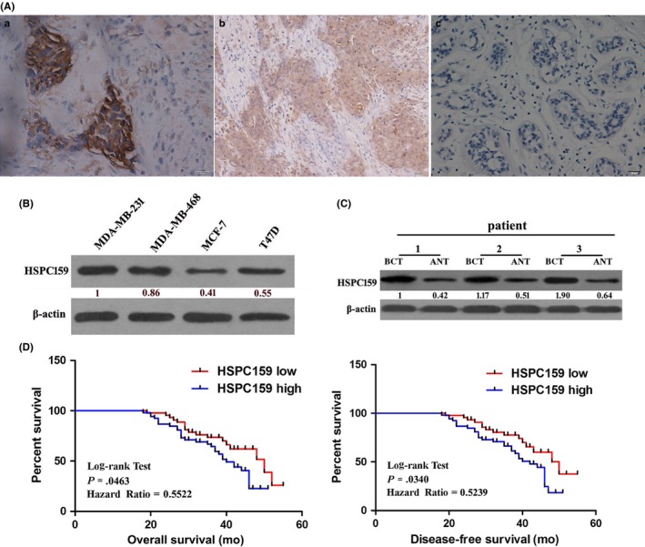
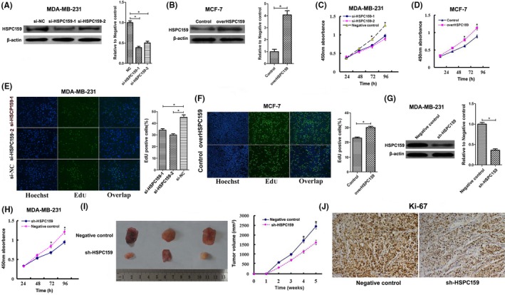
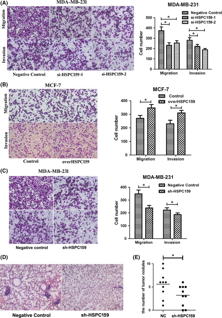
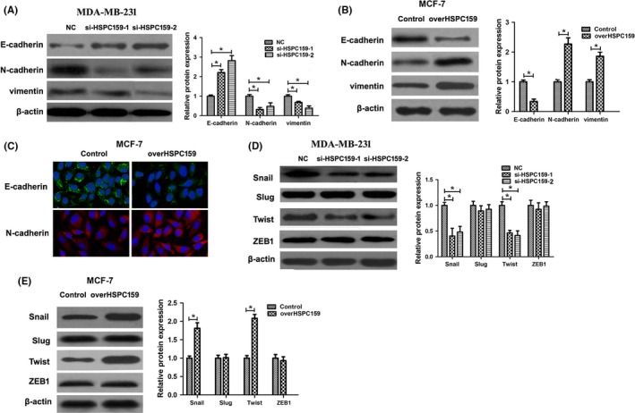
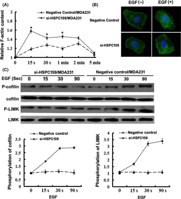
HSPC159 is a novel human galectin-related protein that has been shown to be involved in carcinogenesis. Little is known about HSPC159 expression and function in breast cancer. Herein we showed that HSPC159 was aberrantly expressed in both breast cancer cell lines and tumor tissues and that its expression was associated with poor prognosis of breast cancer patients. Using gain- and loss-of-function methods we found that HSPC159 enhanced breast cancer cell proliferation and metastasis in vitro and in vivo. Mechanistically, HSPC159 was found to induce epithelial-mesenchymal transition (EMT) and the F-actin polymerization process of breast cancer cells. Moreover, HSPC159 promoted proliferation, migration and invasion through activating the PI3K/Akt signaling pathway in breast cancer. In conclusion, our findings showed that HSPC159 contributed to breast cancer progression through the PI3K/Akt pathway and might serve as a potential therapeutic target for the treatment of breast cancer.

Keywords: Akt; EMT; HSPC159; metastasis; proliferation.


Figures

Figure 1
Figure 1
Expression of HSPC159 in breast cancer tissues and invasive breast cancer cell lines. A, Immunohistochemistry analysis of HSPC159 protein levels in 96 primary invasive breast cancer and adjacent normal tissues. a, strong staining of HSPC159 in breast cancer tissues. b, weak staining of HSPC159 in breast cancer tissues. c, negative staining of HSPC159 in adjacent normal tissues. B, Relative expression level of HSPC159 in different breast cancer cell lines. C, HSPC159 protein expression level in paired breast cancer tissues and adjacent non‐cancerous tissues. D, Kaplan‐Meier curves with univariate analysis of overall survival and disease‐free survival based on HSPC159 expression. ANT, adjacent non‐tumor tissues; BCT, human breast cancer tissues
Figure 2
Figure 2
HSPC159 promoted breast cancer cell proliferation in vitro and in vivo. A, Western blot analysis showed siRNA targeting HSPC159 reduced HSPC159 expression level in MDAMB‐231 cells. B, Western blot analysis showed that the protein level of HSPC159 was increased after transfection with pcDNA3.1‐HSPC159 plasmid compared with negative control in MCF‐7 cells. C,D, Cell proliferation rates were measured using CCK‐8 assays in MDAMB‐231 cells (C) and MCF‐7 cells (D) after transfection. E,F, Results of EdU assays also confirmed the effect of HSPC159 on cell proliferation. G, Western blot analysis showed shRNA targeting HSPC159 reduced HSPC159 expression level in MDAMB‐231 cells. H, Cell proliferation rate was measured using CCK‐8 assays in MDAMB‐231 cells after shRNA transfection. I, Knockdown of HSPC159 in MDAMB‐231 cells suppressed tumorigenesis in vivo. Tumor volumes were measured on the indicated days. J, Immunohistochemistry for Ki‐67 detection showed that tumor cells in the negative control group showed a higher positivity rate than those in the shRNAHSPC159 group (magnification ×200). *< .05
Figure 3
Figure 3
HSPC159 enhanced the migration and invasion ability of breast cancer cells in vitro and in vivo. A, Transwell assays showed that depleted cells had less migratory and invasive capacity in MDAMB‐231 cells. B, Effect of HSPC159 overexpression on cell migration and invasion ability in MCF‐7 cells. C, MDAMB‐231 cells showed reduced migration and invasion capacity after shRNA transfection. D, Representative metastasis images are shown with H&E staining. E, Number of lung metastases of indicated SCID mice groups. *< .05
Figure 4
Figure 4
Knockdown of HSPC159 inhibited the epithelial‐mesenchymal transition (EMT) process of breast cancer cells. A, Western blot analysis showed that EMT marker E‐cadherin was upregulated, and that EMT markers N‐cadherin and vimentin were downregulated after knockdown of HSPC159 in MDAMB‐231 cells. B, Western blot analysis showed that EMT marker E‐cadherin was downregulated, and that N‐cadherin and vimentin were upregulated after overexpression of HSPC159 in MCF‐7 cells. C, Immunofluorescence staining of E‐cadherin and N‐cadherin after overexpression of HSPC159 in MCF‐7 cells. D,E, Expression of EMT‐associated transcription factors in MDAMB‐231 and MCF‐7 cells by western blot. *< .05
Figure 5
Figure 5
HSPC159 promoted F‐actin polymerization in breast cancer cells. A, Time course of relative F‐actin content in MDAMB‐231 cells. B, Cytoskeleton rearrangement in si‐HSPC159/MDA231 and negative control cells was imaged by immunofluorescence assay. Cells were treated with 10 ng/mL epidermal growth factor (EGF). Figures show representative images from 3 repeated experiments. C, Western blot analysis of the phosphorylation of cofilin and LIMK in total cell lysates from MDAMB‐231 cells with EGF stimulation for 0, 15, 30 and 90 min. Total cofilin and LIMK was used as a loading control. *< .05
Figure 6
Figure 6
HSPC159 enhanced proliferation, migration and invasion through the PI3K/Akt pathway in breast cancer cells. A,B, Western blot analysis of the phosphorylation of Akt (Thr308 and Ser473) in total cell lysates from MDAMB‐231 and MCF‐7 cells. MDAMB‐231 and MCF‐7 cells were pretreated with PI3K inhibitors LY294002 for 1 h, and the expression level of Akt (Thr308 and Ser473) phosphorylation was detected by western blot. Akt was used as a loading control. C,D, Cell proliferation rates were measured using CCK‐8 (C) and EdU assays (D) after overexpression of HSPC159 with or without LY294002 treatment in MCF‐7 cells. E, Migration and invasion ability was measured by Transwell assays after upregulated expression of HSPC159 with or without LY294002 treatment in MCF‐7 cells. *< .05

References

    1. Torre LA, Bray F, Siegel RL, et al. Global cancer statistics, 2012. CA Cancer J Clin. 2015;65:87‐108. - PubMed
    1. Liu FT, Rabinovich GA. Galectins as modulators of tumour progression. Nat Rev Cancer. 2005;5:29‐41. - PubMed
    1. Wu MH, Hong TM, Cheng HW, et al. Galectin‐1‐mediated tumor invasion and metastasis, up‐regulated matrix metalloproteinase expression, and reorganized actin cytoskeletons. Mol Cancer Res. 2009;7:311‐318. - PubMed
    1. Chen C, Duckworth CA, Fu B, Pritchard DM, Rhodes JM, Yu LG. Circulating galectins ‐2, ‐4 and ‐8 in cancer patients make important contributions to the increased circulation of several cytokines and chemokines that promote angiogenesis and metastasis. Br J Cancer. 2014;110:741‐752. - PMC - PubMed
    1. Zhang QH, Ye M, Wu XY, et al. Cloning and functional analysis of cDNAs with open reading frames for 300 previously undefined genes expressed in CD34+ hematopoietic stem/progenitor cells. Genome Res. 2000;10:1546‐1560. - PMC - PubMed