Atypical posterior scleritis mimicking choroidal melanoma
- PMID: 29738013
- PMCID: PMC6118194
- DOI: 10.15537/smj.2018.5.22130
Atypical posterior scleritis mimicking choroidal melanoma
Abstract
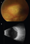
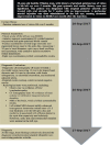
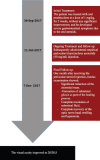
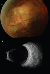
We report a case of atypical posterior scleritis mimicking amelanotic choroidal melanoma. A 30-year-old healthy Filipino man, with a history of painless subacute loss of vision in his left eye over 5 months, was referred to our institute for further workup and management. On examination, visual acuity of the left eye was 20/200. Anterior segment examination yielded unremarkable results, with injected conjunctiva and quiet episcleral blood vessels, while fundus examination revealed non-pigmented nasal choroidal mass, with significant subretinal fluid resembling amelanotic choroidal melanoma. Right eye examination yielded unremarkable results. The patient was diagnosed with atypical posterior scleritis, and treated with oral steroids for 2 weeks, with no improvement. A periocular steroid was then injected to the left eye, causing dramatic reduction in choroidal mass size, and complete resolution of subretinal fluid. The visual acuity improved to 20/28.5 one month after the injection. Timely treatment was crucial for minimizing vision-threatening complications.
Figures


References
-
- Benson WE. Posterior scleritis. Surv Ophthalmol. 1988;32:297–316. - PubMed
-
- Demirci H, Shields CL, Honavar SG, Shields JA, Bardenstein DS. Long-term Follow-up of Giant Nodular Posterior Scleritis Simulating Choroidal Melanoma. Trans R Soc Trop Med Hyg. 1973;67:276. - PubMed
-
- McCluskey Watson PJ, Lightman PG. Posterior scleritis: clinical features, systemic associations, and outcome in a large series of patients. Ophthalmology. 1999;106:2380–1386. - PubMed
-
- Lavric A, Gonzalez-Lopez JJ, Majumder PD, et al. Posterior Scleritis: Analysis of Epidemiology, Clinical Factors, and Risk of Recurrence in a Cohort of 114 Patients. Ocular immunology and Inflammation. 2016;24:6–15. - PubMed
Publication types
MeSH terms
LinkOut - more resources
Full Text Sources
Other Literature Sources
Medical

