. 2018 May 8;19(5):1404.
doi: 10.3390/ijms19051404.

Human Macrophages Preferentially Infiltrate the Superficial Adipose Tissue

Affiliations

Human Macrophages Preferentially Infiltrate the Superficial Adipose Tissue

Giuseppe Cappellano et al. Int J Mol Sci. .

Abstract

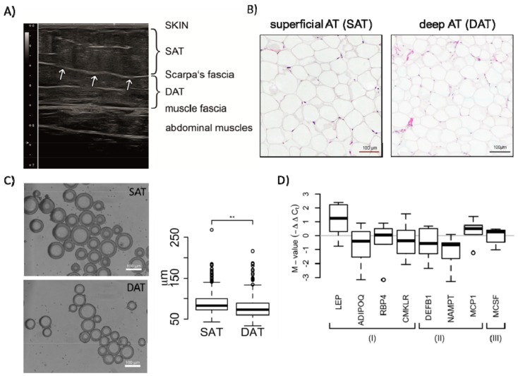
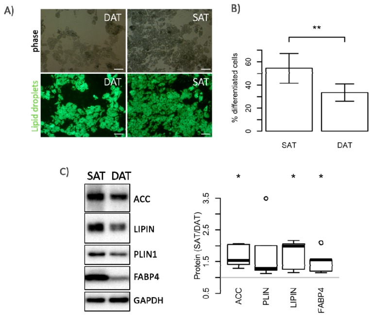
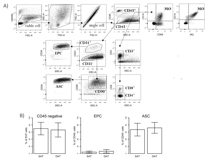
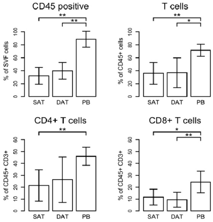
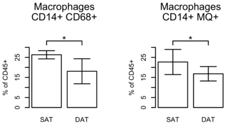
Human abdominal subcutaneous adipose tissue consists of two individual layers—the superficial adipose tissue (SAT) and deep adipose tissue (DAT)—separated by the Scarpa’s fascia. The present study focuses on the analysis of morphological and immunological differences of primary adipocytes, adipose-derived stem cells (ASC), and tissue-infiltrating immune cells found in SAT and DAT. Adipocytes and stromal vascular fraction (SVF) cells were isolated from human SAT and DAT specimens and phenotypically characterized by in vitro assays. Ex vivo analysis of infiltrating immune cells was performed by flow cytometry. Primary adipocytes from SAT are larger in size but did not significantly differ in cytokine levels of LEPTIN, ADIPOQ, RBP4, CHEMERIN, DEFB1, VISFATIN, MCP1, or MSCF. ASC isolated from SAT proliferated faster and exhibited a higher differentiation potential than those isolated from DAT. Flow cytometry analysis indicated no specific differences in the relative numbers of ASC, epithelial progenitor cells (EPC), or CD3⁺ T-cells, but showed higher numbers of tissue-infiltrating macrophages in SAT compared to DAT. Our findings suggest that ASC isolated from SAT have a higher regenerative potential than DAT-ASC. Moreover, spatial proximity to skin microbiota might promote macrophage infiltration in SAT.

Keywords: adipose-derived stem cells; deep adipose tissue; immune cell infiltration; macrophages; superficial adipose tissue.


Conflict of interest statement

The authors declare no conflict of interest.

Figures

Figure 1
Figure 1
Morphological and paracrine characterization of superficial adipose tissue (SAT) and deep adipose tissue (DAT) adipocytes. (A) Representative ultrasound image of infraumbilical subcutaneous fat tissue showing the two individual subcutaneous fat layers. The arrows indicate the Scarpa’s fascia. Obviously, a higher level of hyperechogenic connective tissue structures was observed in DAT indicating structural fat tissue architecture and functional differences; (B) images of H&E-stained SAT and DAT cross-sections; (C) microscopy and quantitative analyses of freshly isolated adipocytes from SAT or DAT. The box plot represents data from a total of 2167 analysed adipocytes isolated from 3 female patients (Student’s t-test, ** p-values < 0.01); (D) RNA from SAT and DAT adipocytes was analysed for the expression of depicted cytokines by quantitative real time PCR. Expression values of indicated cytokines from six patients were normalized to the mean of three reference genes (GUSB, 18sRNA, and GAPDH) and are grouped according their function: (I) represents adipokines, (II) cytokines involved in inflammation and pathogen defence, (III) cytokine associated with neoangiogenesis. Shown are distributions of M-values (log2 fold-change values representing differential expression between SAT and DAT). Significance for difference of the means was calculated using a paired t-test.
Figure 2
Figure 2
Stromal vascular fraction (SVF) cellularity and proliferation capacity of SAT- and DAT-derived adipose-derived stem cells (ASC). (A) Cellularity was calculated by correlating the numbers of SVF cells with the amount (g) of processed fat tissue. Data are shown as mean ± SD (n = 6); (B,C) proliferation of SAT and DAT ASC was assessed after culture for 6 days by analysing DNA content (CyQANT) and mitochondrial activity (PrestoBlue). Results are shown as % of DAT (set to 100%) from six patients; (D) representative immunoblot and quantitative assessment of day 3 proliferating ASC from four donors, analysing expression and phosphorylation of protein kinase B (AKT), extracellular signal-regulated kinase ERK 1/2 (p44/42), mammalian target of rapamycin (mTOR), and GAPDH as loading control. Data are shown as mean ± SD. Significance for difference of the means was calculated using a paired t-test (* p-value < 0.05).
Figure 3
Figure 3
Adipocyte differentiation potential of SAT- and DAT-derived ASC. ASC isolated from SAT and DAT were differentiated in vitro for 14 days. BODIPY™ 493/503-stained adipocytes were analysed by fluorescence microscopy (A) and quantitatively assessed by flow cytometry (B); Size bar: 100 µm. Data are shown as mean ± SD (n = 6), significance for difference of the means calculated with a paired t-test (** p-value < 0.01); (C) Representative immunoblot and quantitative assessment of day 14 differentiated ASC from five donors, analysing expression of acetyl-CoA carboxylase (ACC), lipid-droplet-binding protein perilipin 1 (PLIN1), phosphatidate phosphatase lipin 1 (LPIN1), fatty acid transport protein 4 (FABP4), and GAPDH as loading control. Results are shown as box plots representing the distribution of fold change values; significance of the fold change was assessed by testing against the null hypothesis of a mean fold change of 1 (* p-value < 0.05).
Figure 4
Figure 4
Flow cytometry analysis of ASC, endothelial progenitor cells (EPC), and T-cells in SVF from SAT, DAT, and blood. SVF cells isolated from SAT and DAT as well as peripheral blood mononuclear cells (PBMC) from paired samples were analysed by flow cytometry. (A) Representative FACS plots showing the gating strategy of different cell populations investigated in this study (FSC-A: forward scatter area; SSC-A: side scatter area; FSC-H: forward scatter height); (B) percentages of CD45+ and CD45 are shown on viable cells. For further analysis, the percentages of cells were calculated based on CD45+ and CD45 cells, respectively. EPC (CD45CD31+CD34+) and ASC (CD45CD31CD90+CD34+) are shown as percentage (%) of CD45 cells. Results represent data from five patients and are expressed as mean ± SD.
Figure 5
Figure 5
Analysis of T-cells in SAT, DAT, and peripheral blood cells (PB). Gating strategy is shown in Figure 4A. The percentages of T-cells were calculated based on the numbers of CD45+ cells. CD8+ T-cells were discriminated from CD4+ T-helper cells on the basis of expression of CD8 marker. CD4+ T-cells were determined as CD8- cells. Results represent data from six patients and are expressed as mean ± SD. Significance was assessed using a paired t-test (* p-value < 0.05, ** p-value < 0.01).
Figure 6
Figure 6
Macrophage infiltration in SAT and DAT. Gating strategy is shown in Figure 4A. Macrophages (defined as CD14+CD68+ or CD14++ (clone 25f9)) are shown as % of CD45+ cells. Results represent data from four patients and are expressed as mean ± SD. Significance of the difference in means was calculated using a paired t-test (* p-value < 0.05).

References

    1. McLaughlin T., Lamendola C., Liu A., Abbasi F. Preferential fat deposition in subcutaneous versus visceral depots is associated with insulin sensitivity. J. Clin. Endocrinol. Metab. 2011;96:E1756–E1760. doi: 10.1210/jc.2011-0615. - DOI - PMC - PubMed
    1. Wronkowitz N., Romacho T., Sell H., Eckel J. Adipose tissue dysfunction and inflammation in cardiovascular disease. Front. Horm. Res. 2014;43:79–92. doi: 10.1159/000360560. - DOI - PubMed
    1. Lovren F., Teoh H., Verma S. Obesity and atherosclerosis: Mechanistic insights. Can. J. Cardiol. 2015;31:177–183. doi: 10.1016/j.cjca.2014.11.031. - DOI - PubMed
    1. Driskell R.R., Jahoda C.A., Chuong C.M., Watt F.M., Horsley V. Defining dermal adipose tissue. Exp. Dermatol. 2014;23:629–631. doi: 10.1111/exd.12450. - DOI - PMC - PubMed
    1. Nakajima H., Imanishi N., Minabe T., Kishi K., Aiso S. Anatomical study of subcutaneous adipofascial tissue: A concept of the protective adipofascial system (PAFS) and lubricant adipofascial system (LAFS) Scand. J. Plast. Reconstr. Surg. Hand Surg. 2004;38:261–266. doi: 10.1080/02844310410029543. - DOI - PubMed

MeSH terms