. 2018 Sep 1;24(17):4201-4214.
doi: 10.1158/1078-0432.CCR-18-0410. Epub 2018 May 8.

MAPK Reliance via Acquired CDK4/6 Inhibitor Resistance in Cancer

Affiliations

MAPK Reliance via Acquired CDK4/6 Inhibitor Resistance in Cancer

Renée de Leeuw et al. Clin Cancer Res. .

Abstract

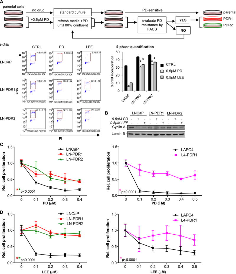
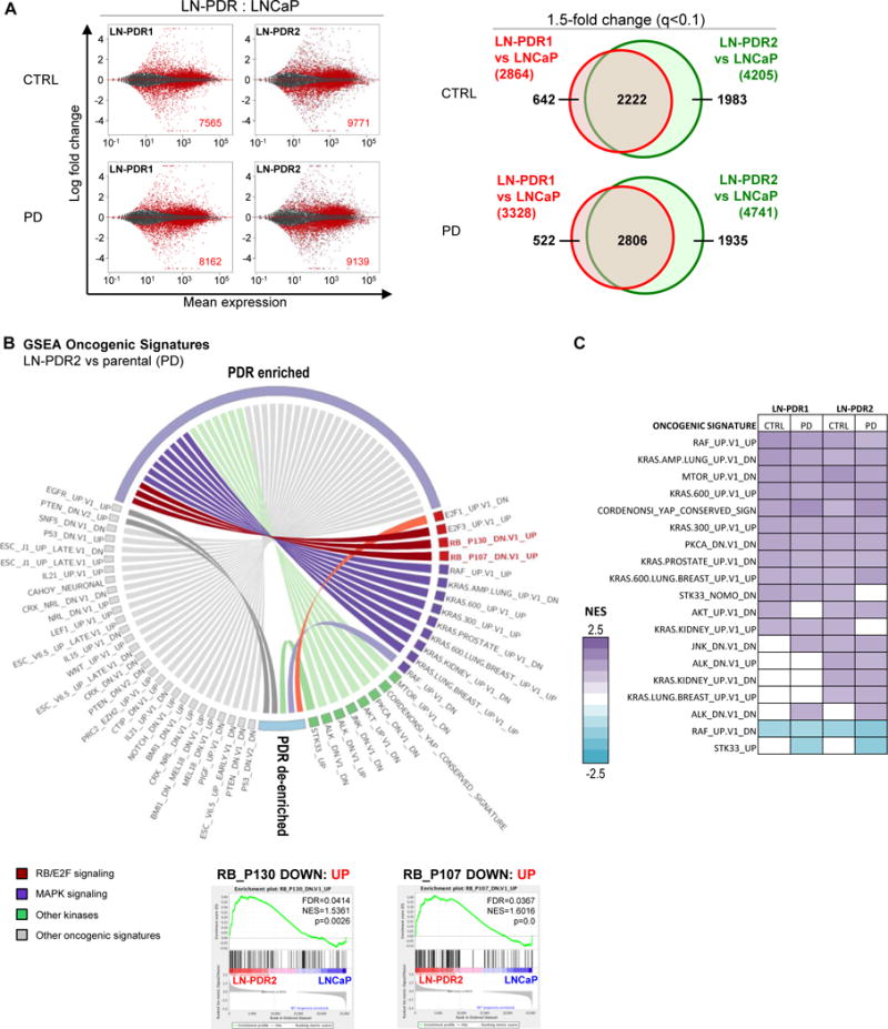
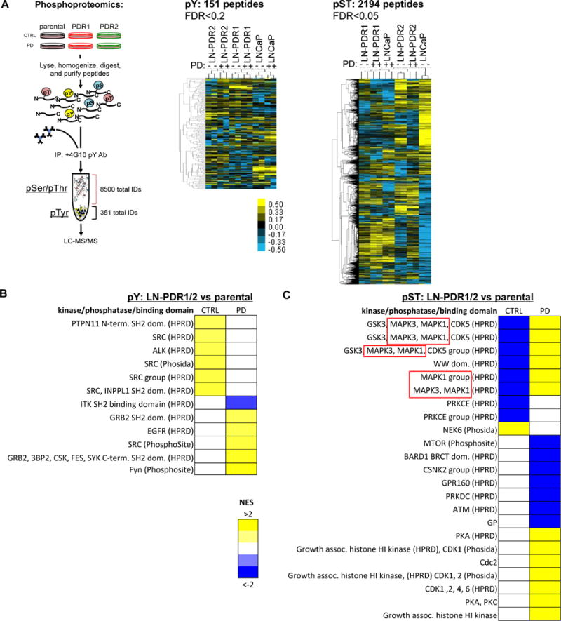
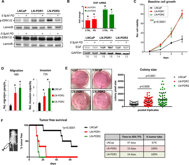
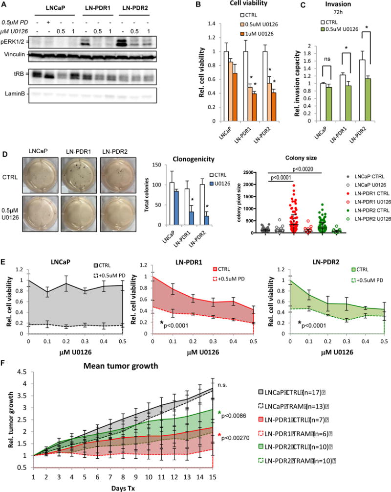
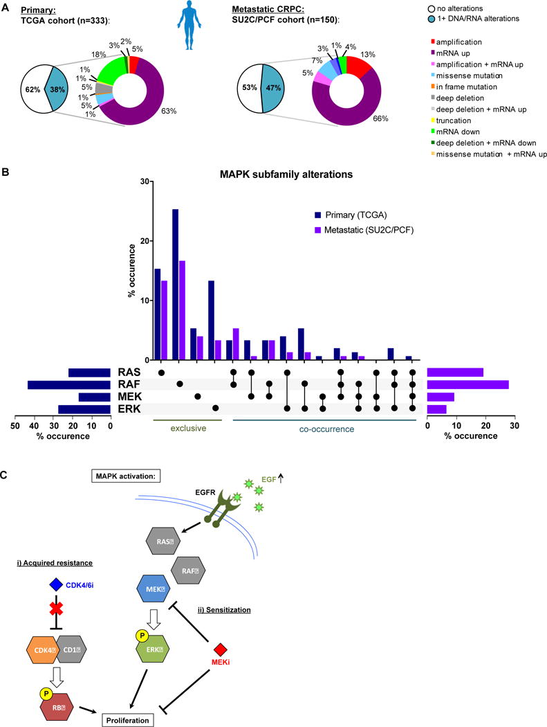
Purpose: Loss of cell-cycle control is a hallmark of cancer, which can be targeted with agents, including cyclin-dependent kinase-4/6 (CDK4/6) kinase inhibitors that impinge upon the G1-S cell-cycle checkpoint via maintaining activity of the retinoblastoma tumor suppressor (RB). This class of drugs is under clinical investigation for various solid tumor types and has recently been FDA-approved for treatment of breast cancer. However, development of therapeutic resistance is not uncommon.Experimental Design: In this study, palbociclib (a CDK4/6 inhibitor) resistance was established in models of early stage, RB-positive cancer.Results: This study demonstrates that acquired palbociclib resistance renders cancer cells broadly resistant to CDK4/6 inhibitors. Acquired resistance was associated with aggressive in vitro and in vivo phenotypes, including proliferation, migration, and invasion. Integration of RNA sequencing analysis and phosphoproteomics profiling revealed rewiring of the kinome, with a strong enrichment for enhanced MAPK signaling across all resistance models, which resulted in aggressive in vitro and in vivo phenotypes and prometastatic signaling. However, CDK4/6 inhibitor-resistant models were sensitized to MEK inhibitors, revealing reliance on active MAPK signaling to promote tumor cell growth and invasion.Conclusions: In sum, these studies identify MAPK reliance in acquired CDK4/6 inhibitor resistance that promotes aggressive disease, while nominating MEK inhibition as putative novel therapeutic strategy to treat or prevent CDK4/6 inhibitor resistance in cancer. Clin Cancer Res; 24(17); 4201-14. ©2018 AACR.


Conflict of interest statement

COI: The authors declare no potential conflicts of interest.

Figures

Figure 1
Figure 1. Acquired resistance to palbociclib results in broad CDK4/6 inhibitor resistance
A. Palbociclib (PD) resistant (PDR) prostate cancer cells were generated via continuous selection with 0.5μM PD for 2-3 months and evaluated regularly via flow cytometry (top). PD resistance was determined by treating biological triplicate parental or PDR cells with 0.5μM PD, LEE (ribociclib), or CTRL (no drug) for 24h and measuring BrdU incorporation with a flow cytometer after a 2 hour pulse labeling, fixation in EtOH and staining with a secondary FITC-mouse-anti-BrdU antibody. FACS analysis was performed by gating for the BrdU+ S-phase population (representative flow traces for biological triplicates are shown on the left), quantified in a bar graph on the right as an indication of cell proliferation. FACS analysis showed that PD and ribociclib (LEE) fail to induce cell cycle arrest in the G1-phase in LNCaP PDR lines. B. Cells were treated for 24h with CTRL, 0.5μM PD, or LEE and immunoblotted for Cyclin A, demonstrating a reduction in Cyclin A protein upon exposure to PD or LEE only in the parental cells, indicating biochemical resistance in LNCaP PDR cells. C. Cell counting via Trypan blue exclusion of quadruplet samples with dose escalation treatment with PD of t=6 days (LNCaP) or t=13 days (LAPC4) shows a significantly reduced response to PD in the PDR models, compared to the parental cells. D. Acquired resistance to PD results in broad CDK4/6 inhibitor resistance as shown by a LEE dose escalation. *Significance for dose response curves was determined by a Two-way ANOVA analysis performing a multivariate comparison of mean per dose for PDR vs parental data.
Figure 2
Figure 2. Acquired CDK4/6 inhibitor resistance is associated with rewired transcriptional programs including RB function
A. RNAseq was performed on PDR1/2 and parental LNCaP cells treated 24h with 0.5μM PD or vehicle (CTRL). The MA plots (right) represent the log ratio (M) of PDR versus parental values over the average log intensity (A) of each transcript, which visualizes vast differences between the PDR vs parental data (red dots and inset numbers indicate significant hits, q-value<0.1). Venn diagrams show overlap between PDR1 and PDR2 of genes >1.5x differentially expressed compared to the parental cells (q-value<0.1; right). B. The complete RNAseq profiles for each PDR vs parental model comparison were subjected to unbiased Gene Set Enrichment Analysis (GSEA, MSigDB) to determine enrichment of predefined Oncogenic Signatures in both PDR1 and PDR2 compared to parental cells under at least one condition (CTRL/PD) with a False Discovery Rate or FDR<0.25 (Complete list in Supplementary Figure 2B). The Oncogenic Signatures enriched in the PDR models included two signatures defined by RB knockdown, suggesting the PDR models have upregulated genes that are induced by RB knockdown. Representative GSEA plots of the RB knockdown signatures are shown for PDR2 vs WT after PD treatment. C. GSEA Oncogenic Signature altered kinase signatures in the LNCaP PDR models for all conditions.
Figure 3
Figure 3. Integrative transcriptome and kinome profiling identifies differential MAPK stimulus as a hallmark of CDK4/6 inhibitor resistance
A. LNCaP PDR and parental cells were treated for 24h with 0.5uM PD or control, snap frozen and lysed. After peptide digestion, phospho-Tyrosine peptides (pY) were immunoprecipitated, while phospho-Serine/Threonine peptides (pST) remained in the upper fraction (see schematic, more details in Materials and Methods). Duplicates of both peptide fractions were utilized in an unbiased phosphoproteomics approach to identify altered phosphorylation of Tyrosine and Serine/Threonine peptides across PDR models compared to the parental cells, displayed by hierarchical clustering on the right. B. Kinase/Substrate Enrichment Analysis (KSEA) defined enriched peptide motifs for phosphorylated Tyrosine (pY) hits and mapped them to kinases that are most likely to target these motifs. This revealed enrichment for Src and Src Homology (SH2) domain target motifs in PDR1/2 compared to parental cells (p=0.2). C. KSEA analysis for pST hits showed enrichment for altered phosphorylation of MAPK3 and MAPK1 (ERK1/2) target motifs (p=0.05), indicative of differential MAPK signaling.
Figure 4
Figure 4. Acquired CDK4/6 inhibitor resistance promotes aggressive phenotypes
A. LNCaP PDR and parental cells were treated for 24h with 0.5μM PD or CTRL, lysed and immunoblotted for phospho-ERK1/2 (P-ERK1/2) and total ERK1/2 (t-ERK1/2). Results show hyperphosphorylation of ERK1/2 in the PDR lines compared to parental cells, while total protein levels are unchanged across the different models and conditions. These data indicate that the MAPK pathway is activated in the PDR models. B. EGF transcript level fold changes of LNCaP PDR1/2 relative to parental from RNAseq data (top, *q-value<0.1; error bars: standard error) show elevated EGF mRNA, resulting in increased EGF protein expression via Western blotting (bottom, numbers represent EGF quantification normalized to GAPDH, relative to parental CTRL. C. A time course experiment to assess cell proliferation via Trypan blue exclusion revealed that the PDR cells off PD selection are hyperproliferative compared to parental cells (*Significance was determined by a Two-way ANOVA analysis performing a multivariate comparison of mean per time point for PDR vs parental; p<0.0001). D. Cells were seeded in 6 replicates in serum-free media in a Fluoroblok transwell migration (without matrigel) or invasion (with matrigel) plates and allowed to migrate for 48h or invade for 72h to serum-rich (20%) media in the bottom of the well. PDR cells display enhanced migratory capacity and invasion through matrigel (*Student’s t-test: p<0.05 vs parental). E. Clonogenic capacity of the PDR and parental lines was assessed by seeding cells at low density (5,000/well in a 6-wells plate) in media supplemented with 0.6% agar and left to grow 3 weeks. The PDR lines displayed the ability to grow larger 3D colonies in agar. Images (left) show representative wells of triplicates. “+ctrl” are MAF cells with known clonogenicity. Colony sizes were determined via image pixel counts in FIJI (see Materials and Methods), triplicates were pooled and plotted (right) as single dots according to size (Y-axis). Line and whiskers show median and 95% Confidence Interval (CI, *Statistical significance determined by a One-way ANOVA compared to parental). F. Sub-cutaneous xenograft tumor growth of LNCaP PDR and parental cells injected at 3×10⁶ cells in the flanks of athymic nude mice (n=6 per group) reveal accelerated tumor formation in vivo, graphed as % tumor free survival (TFS) over time (left). *Statistical significance determined via One-way ANOVA compared to parental. Table (right) shows reduced time to 50% tumor free survival (TFS) and enhanced overall tumor take.
Figure 5
Figure 5. CDK4/6i resistant models become reliant on activated MAPK pathway and sensitized to MEK inhibition
A. LNCaP cells were treated 24h with 0.5 or 1μM MEK inhibitor (U0126) or CTRL and immunoblotted for p-ERK1/2, demonstrating loss of the hyperphosphorylation of ERK1/2 observed in the LNCaP PDR models when MEK is inhibited, confirming that the MAPK pathway is activated in the PDR models. MEK inhibition does not affect RB hyperphosphorylation (upper band tRB) or CDK4 phosphorylation, indicating a bypass of the RB cell cycle checkpoint. B. Cell counting via Trypan blue exclusion after 6 days of 0.5 or 1μM U0126 treatment reveals that the PDR models are sensitized to MEK inhibition (*significant difference compared to corresponding treatment in parental cells). C. Invasion of PDR cells, but not parental cells, through matrigel in a Fluoroblok transwell system decreases with a MEK inhibitor (*p<0.05 vs parental). D. Clonogenic assay shows reduction in both size (center) and total numbers of colonies (right) formed by the PDR models, whereas the parental cells are unresponsive to MEK inhibition (0.5μM U0128). E. Cell counting with escalating doses of U0126 (0-0.5uM, t=6d) shows a cooperative effect with 0.5uM PD in PDR cells. F. LNCaP parental and PDR cells were injected sub-Q into the flanks of athymic nude mice. When tumors reached ~150mm3, mice were treated with chow laced with trametinib (TRAM). Caliper measurements revealed that, while the parental tumors were unresponsive to the MEK inhibitor,, the PDR models are sensitized to trametinib in vivo (error bars represent SEM).
Figure 6
Figure 6. cBioportal analysis of clinical datasets reveals MAPK subfamily alterations in primary and metastatic tumors
A. cBioportal analyses of the TCGA clinical cohort shows alterations in DNA and/or RNA for MAPK pathway genes in 128/333 primary prostate cancer patients (38%). Similarly, SU2C/PCF cohort reveals alterations in 71/150 patients with advanced prostate cancer (~47%). The majority of alterations in these patient datasets are gene amplification and/or transcriptional upregulation (~68% and ~85%, respectively). B. MAPK subfamily kinase analysis of alterations observed in primary (blue) and metastatic (purple) showing total percentages per subfamily (horizontal bar graphs), and exclusive versus co-occuring subfamily alterations (vertical bar graph, black dots indicate alteration, details per patient in Supplementary Figure 7). C. Schematic of acquired CDK4/6 inhibitor resistance. The PDR models presented here have acquired resistance to CDK4/6 inhibition, resulting in proliferation and aggressive phenotypes. This acquired resistance is associated with MAPK pathway activation, creating a delicate reliance on this pathway that is independent of the RB cell cycle checkpoint. This MAPK activation resulted in sensitization to MEK inhibition. EGF upregulation was observed in the PDR models, which activates the EGF Receptor (EGFR), and likely activates MAPK signaling downstream. While CDK4/6 inhibitors fail to block tumor growth in these models of acquired resistance, the sensitization to MEK inhibition provides new rationale for treating cancers that have acquired resistance to CDK4/6 inhibitors.

References

    1. Barroso-Sousa R, Shapiro GI, Tolaney SM. Clinical Development of the CDK4/6 Inhibitors Ribociclib and Abemaciclib in Breast Cancer. Breast Care (Basel) 2016;11:167–173. - PMC - PubMed
    1. Tripathy D, Bardia A, Sellers WR. Ribociclib (LEE011): mechanism of action and clinical impact of this selective cyclin-dependent kinase 4/6 inhibitor in various solid tumors. Clinical cancer research : an official journal of the American Association for Cancer Research. 2017 - PMC - PubMed
    1. Garrido-Castro AC, Shom Goel S. CDK4/6 Inhibition in Breast Cancer: Mechanisms of Response and Treatment Failure. Curr Breast Cancer Rep. 2017:26–33. - PMC - PubMed
    1. Vijayaraghavan S, Moulder S, Keyomarsi K, Layman RM. Inhibiting CDK in Cancer Therapy: Current Evidence and Future Directions. Target Oncol. 2017 - PubMed
    1. Comstock CE, Augello MA, Goodwin JF, de Leeuw R, Schiewer MJ, Ostrander WF, Jr, et al. Targeting cell cycle and hormone receptor pathways in cancer. Oncogene. 2013;32:5481–5491. - PMC - PubMed

Publication types

MeSH terms