. 2018 May 8;9(3):e00625-18.
doi: 10.1128/mBio.00625-18.

Bone Marrow Is a Major Parasite Reservoir in Plasmodium vivax Infection

Affiliations

Bone Marrow Is a Major Parasite Reservoir in Plasmodium vivax Infection

Nicanor Obaldia 3rd et al. mBio. .

Abstract

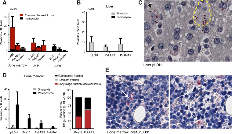
Plasmodium vivax causes heavy burdens of disease across malarious regions worldwide. Mature P. vivax asexual and transmissive gametocyte stages occur in the blood circulation, and it is often assumed that accumulation/sequestration in tissues is not an important phase in their development. Here, we present a systematic study of P. vivax stage distributions in infected tissues of nonhuman primate (NHP) malaria models as well as in blood from human infections. In a comparative analysis of the transcriptomes of P. vivax and Plasmodium falciparum blood-stage parasites, we found a conserved cascade of stage-specific gene expression despite the greatly different gametocyte maturity times of these two species. Using this knowledge, we validated a set of conserved asexual- and gametocyte-stage markers both by quantitative real-time PCR and by antibody assays of peripheral blood samples from infected patients and NHP (Aotus sp.). Histological analyses of P. vivax parasites in organs of 13 infected NHP (Aotus and Saimiri species) demonstrated a major fraction of immature gametocytes in the parenchyma of the bone marrow, while asexual schizont forms were enriched to a somewhat lesser extent in this region of the bone marrow as well as in sinusoids of the liver. These findings suggest that the bone marrow is an important reservoir for gametocyte development and proliferation of malaria parasites.IMPORTANCEPlasmodium vivax malaria continues to cause major public health burdens worldwide. Yet, significant knowledge gaps in the basic biology and epidemiology of P. vivax malaria remain, largely due to limited available tools for research and diagnostics. Here, we present a systematic examination of tissue sequestration during P. vivax infection. Studies of nonhuman primates and malaria patients revealed enrichment of developing sexual stages (gametocytes) and mature replicative stages (schizonts) in the bone marrow and liver, relative to those present in peripheral blood. Identification of the bone marrow as a major P. vivax tissue reservoir has important implications for parasite diagnosis and treatment.

Keywords: Aotus; Saimiri; blood-stage parasites; gametocytes; immunohistochemistry; laboratory animal models; malaria; real-time PCR; transcriptome.


Figures

FIG 1
FIG 1
Stage-specific P. vivax gene expression ex vivo and in patients. (A) P. vivax gametocyte transcriptional dynamics ex vivo. P. vivax ex vivo transcriptome data from three patient isolates (31) were reanalyzed for gametocyte transcriptional dynamics. In the heat map, P. vivax orthologs from P. falciparum gametocyte genes were sorted along the developmental cycle according to stage specificity of previously defined clusters of P. falciparum coexpression analysis (29) (see Materials and Methods). The progression of transcript orthologs through the stages of P. vivax and P. falciparum gametocyte development was similar, although the period of the immature gametocyte cycle was greatly extended in P. falciparum compared to P. vivax. (B) Three selected transcriptional clusters including P. vivax markers PvPPM3 (cluster 240), PvLAP5 (cluster 241), and Pvg377 and Pvs25 (both cluster 49). (C) P. vivax gene expression in patient samples. P. vivax transcriptome data from 8 patient isolates (CM12, CM13, CM101, CM106, CM108, CM114, CM115, and CMM08) (34) were reanalyzed to define gametocyte transcript abundance in patient peripheral blood. (Left) Box plot showing mean expression across all genes representing one of five categories: A. circ, asexual 0 to 22 hpi; A. seq., 22 to 48 hpi (gametocyte GR, IG, and MG). ***, P < 0.005, paired t test. (Right) Heat map showing mean expression per cluster (sorted as for panel A) for GR, IG, and MG stages. (Bottom) Heat map showing mean expression of young circulating asexual stages and more mature asexual stages which accumulate/sequester in tissues. Dashed lines demarcate categories defined previously for P. falciparum (29).
FIG 2
FIG 2
Validation of stage-specific P. vivax markers. (A) P. vivax infection dynamics in Aotus. Samples were collected every 3 to 5 days starting at peak parasitemia and analyzed by qRT-PCR and microscopy. qRT-PCR data were normalized to RNA input and presented as transformed cycle threshold (CT) values. (B) qRT-PCR quantification of candidate markers in the peripheral blood and corresponding 48-h ex vivo samples from Aotus monkeys infected with P. vivax AMRU-I or SAL-I strains (n = 3). Candidate markers and Pvs25 were quantified by qRT-PCR, and values were normalized using PVX18s rRNA. Data are presented as transformed CT values. (C) Validation of marker antibodies by Western blotting using Percoll-enriched blood samples from Aotus infected with P. vivax SAL-I. (Left) A single band was detected in the SAL-I Aotus lysate when we used mouse monoclonal pLDH antibody at a 1:5,000 dilution (approximately 1 × 10⁶ parasites/lane). (Middle) Rabbit anti-PvLAP5 peptide antibodies (1:1,000 dilution) detected full-length protein at 98.2 kDa (arrow) in lysates from infected Aotus (lanes 1 and 3) and the P. falciparum ortholog at 100 kDa in lysate of the P. falciparum HB3 line (lane 4, approximately 1 × 10⁶ parasites/lane from cultivated gametocytes). Lane 2, empty. (Right) Lane 1, Aotus lysate; lane 2, P. falciparum HB3 lysate from schizont cultures. PvAMA1 peptide antibodies (1:1,000) detected a major band and known AMA1 breakdown products in the P. falciparum HB3 schizont lysate but not in Aotus samples, presumably due to low yield (lane 1). (D) Image of Giemsa-stained thin blood film from Aotus ex vivo culture (44 h) showing a P. vivax gametocyte on the left and a vacuolated mature form on the right (arrows). (E, left) IFA images obtained with PvLAP5 (top) and PvAMA1 antibodies (bottom). (Right) Gametocyte percentages determined from PvLAP5 IFA-positive relative to total Giemsa-stained parasite counts in the ex vivo samples from three Brazilian P. vivax patients.
FIG 3
FIG 3
P. vivax tissue accumulation/sequestration in nonhuman primates. (A) Representative images of parasites in the immunohistochemistry (IHC) analysis of 4 tissues. pLDH, PvLAP5, and PvAMA1 antibodies were used to detect the parasite; CD31 antibodies stained the endothelium. Black arrowheads mark parasites. (B) Quantification of histological data. IHC analysis across 6 tissues from 13 monkeys was performed using parasite antibodies against pLDH, PvLAP5, and PvAMA1. Highest counts for all three antibodies were detected in bone marrow, liver, and lung. Counts represent 500 high-power fields. Values are expressed as means ± standard errors of the means (SEM). (Bottom) Pie charts showing parasite distribution across tissues based on PvLAP5 (left) and PvAMA1 counts (right). Parasites in Kupffer cells (liver) were excluded from counts. BM, bone marrow. (C) Parasite and gametocyte burden in blood and tissues. Numbers were calculated based on parasite counts from Giemsa-stained peripheral blood smears and from BM, liver, and lung tissues stained with antibodies for pLDH (all parasites) or PvLAP5 (gametocytes). (Top) Total parasite load in each tissue. (Middle) Total gametocyte load in each tissue. (Bottom) Total schizont load in each tissue. Parasites in Kupffer cells (liver) were excluded from thce ounts. Values are expressed as means ± SEM.
FIG 4
FIG 4
P. vivax accumulation/sequestration in bone marrow and liver. (A) Intra- and extravascular parasite distributions across tissues. Quantification of IHC data across three tissues was based on CD31 staining to differentiate intravascular from extravascular parasite localization. Parasites in macrophages were excluded from the analysis. Data demonstrated that most parasites are extravascular in the bone marrow and liver, but not in the lung. Data are from the tissues of 13 monkeys. mϕ, macrophage. (B) Extravascular parasite distribution in liver. Quantification of IHC data in liver sinusoids and parenchymas is shown. All parasites counted were present in sinusoids. Data are from the tissues of 13 monkeys. (C) Representative image of infected liver tissue. Shown are parasites in Kupffer cells (black arrowhead) and in sinusoids (yellow boundary). (D) IHC results showing extravascular parasite distribution in the bone marrow sinusoids and parenchyma. The majority of parasites counted were in the parenchyma. In bone marrow, gametocytes but not schizonts were mostly extravascular. (Right) Stage fraction in BM parenchyma (stacked bar with pLDH [taken as 100]). Schizonts were quantified based on PvAMA1-positive parasites, and gametocytes were quantified based on Pvs16- or PvLAP5-positive parasites. The remainder (after subtracting PvAMA1 and Pvs16/PvLAP5 counts from pLDH counts) were cataloged as the early-stage fraction. Data are from the tissues of 13 monkeys. (E) Representative images of infected bone marrow tissue. Extravascular parasites are marked with white arrows; the image on the right shows two parasites associated with erythroblastic islands.

Comment in

References

    1. World Health Organization Global Malaria Programme 2015. World malaria report 2015. WHO, Geneva, Switzerland.
    1. Guerra CA, Howes RE, Patil AP, Gething PW, Van Boeckel TP, Temperley WH, Kabaria CW, Tatem AJ, Manh BH, Elyazar IR, Baird JK, Snow RW, Hay SI. 2010. The international limits and population at risk of Plasmodium vivax transmission in 2009. PLoS Negl Trop Dis 4:e774. doi:10.1371/journal.pntd.0000774. - DOI - PMC - PubMed
    1. Bousema T, Drakeley C. 2011. Epidemiology and infectivity of Plasmodium falciparum and Plasmodium vivax gametocytes in relation to malaria control and elimination. Clin Microbiol Rev 24:377–410. doi:10.1128/CMR.00051-10. - DOI - PMC - PubMed
    1. Baird JK. 2013. Evidence and implications of mortality associated with acute Plasmodium vivax malaria. Clin Microbiol Rev 26:36–57. doi:10.1128/CMR.00074-12. - DOI - PMC - PubMed
    1. Zimmerman PA. 2017. Plasmodium vivax infection in Duffy-negative people in Africa. Am J Trop Med Hyg 97:636–638. doi:10.4269/ajtmh.17-0461. - DOI - PMC - PubMed

Publication types

Substances