. 2018 May 8;8(1):7108.
doi: 10.1038/s41598-018-25487-5.

Patient-derived mutations within the N-terminal domains of p85α impact PTEN or Rab5 binding and regulation

Affiliations

Patient-derived mutations within the N-terminal domains of p85α impact PTEN or Rab5 binding and regulation

Paul Mellor et al. Sci Rep. .

Abstract

The p85α protein regulates flux through the PI3K/PTEN signaling pathway, and also controls receptor trafficking via regulation of Rab-family GTPases. In this report, we determined the impact of several cancer patient-derived p85α mutations located within the N-terminal domains of p85α previously shown to bind PTEN and Rab5, and regulate their respective functions. One p85α mutation, L30F, significantly reduced the steady state binding to PTEN, yet enhanced the stimulation of PTEN lipid phosphatase activity. Three other p85α mutations (E137K, K288Q, E297K) also altered the regulation of PTEN catalytic activity. In contrast, many p85α mutations reduced the binding to Rab5 (L30F, I69L, I82F, I177N, E217K), and several impacted the GAP activity of p85α towards Rab5 (E137K, I177N, E217K, E297K). We determined the crystal structure of several of these p85α BH domain mutants (E137K, E217K, R262T E297K) for bovine p85α BH and found that the mutations did not alter the overall domain structure. Thus, several p85α mutations found in human cancers may deregulate PTEN and/or Rab5 regulated pathways to contribute to oncogenesis. We also engineered several experimental mutations within the p85α BH domain and identified L191 and V263 as important for both binding and regulation of Rab5 activity.


Conflict of interest statement

The authors declare no competing interests.

Figures

Figure 1
Figure 1
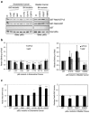
Binding and regulation of PTEN by p85α patient-derived SH3 and BH domain mutants. (a) Schematic representation of p85α with the locations of endometrial and bladder cancer-associated mutations within the SH3 and BH domains shown. (b) Pull-down assays for p85α mutants binding to PTEN. Input of p85α is 4% of the amount used in the pull-down binding experiments. Data representative of at least 9 independent experiments. Full-length images of the cropped blots are in Supplementary Fig. S4. (c) Quantification of binding assay results from panel b. Mean ± SEM. ***P < 0.001 as compared to wild type p85α. (d) The ability of each p85α mutant protein to regulate PTEN was determined using a PTEN lipid phosphatase activity assay. Mean ± SEM for 4–5 independent experiments. ***P < 0.001, **P < 0.01, *P < 0.05 as compared to wild type p85α.
Figure 2
Figure 2
Binding and regulation of Rab5 by p85α patient-derived SH3 and BH domain mutants. (a) Pull-down assay with GST and GST-Rab5 mutants immobilized on glutathione Sepharose beads and loaded with the indicated nucleotide. GTPγS is a non-hydrolyzable analogue of GTP. Binding of purified p85α wild type or mutant protein was detected using an immunoblot analysis. Input of p85α is 4% of the amount used in the pull-down binding experiments. Data representative of at least 4 independent experiments. Full-length images of the cropped blots are in Supplementary Fig. S5. (b) Quantification of binding assay results from panel a. Mean ± SEM. ***P < 0.001, **P < 0.01 as compared to wild type p85α. (c) Rab5 was loaded with [α-32P]-GTP and analyzed for its GTPase activity either alone (no p85α) or in the presence of the indicated p85α protein to measure Rab5 GAP activity. Mean ± SEM for 3 independent experiments. ***P < 0.001, **P < 0.01, *P < 0.05 as compared to wild type p85α.
Figure 3
Figure 3
Crystal structures for bovine p85α (105–319) wild type and containing patient-derived mutations. (a) Overlay of the bovine p85α (105–319) crystal (green and cyan, resolution 2.25 Å) with the crystal structure for the human p85α (105–319) protein fragment (1PBW; magenta and pink, resolution 2.0 Å). A homodimer of the bovine p85α BH domains was visible containing residues 113–297 for both components of the dimer. (b,c) p85α BH domain residues involved in the hydrophobic dimerization interface within the crystal lattice of the bovine protein (b; L161, M176, F177 and V181) and the human protein (c; L161, M176, I177 and V181). (dg) Overlays of the crystal structures for p85α (105–319); wild type (green) and cancer-associated point mutants (yellow): E137K mutant (d), E217K mutant (e), R262T mutant (f), E297K mutant (g). Wild type and mutant sidechains are shown in stick representation. Lack of density for the K297 sidechain prevented inclusion of the sidechain, and it is modeled as Ala in the structure (e).
Figure 4
Figure 4
Residues L191 and V263 in the p85α BH domain are important for Rab5 binding. (a) The BH domain of human p85α with the proposed G protein binding (i.e. Rab5) residues indicated. Residues with little or no effect on Rab5 binding are shown in grey (K187, I267 [L267 in bovine p85α], M271 [L271 in bovine p85α]), with catalytically important R151 in orange. Residues important for both Rab5 binding and catalytic activity are shown in red (L191, V263, R274). Residues that help mediate BH–BH domain dimerization within the crystal structure are shown in M176 (purple) from one BH domain fitting into a hydrophobic pocket containing L161, I177 and V181 (pink) on the other BH domain. (b) Pull-down assay with GST and GST-Rab5 mutants immobilized on glutathione Sepharose beads and loaded with the indicated nucleotide. Binding of purified p85α wild type or mutant protein was detected using an immunoblot analysis. The input lanes contain 0.4% of the purified p85α protein used in the pull-down assay. Full-length images of the cropped blots are in Supplementary Fig. S6. (c) The ability of each p85α mutant protein to regulate Rab5 GTPase activity was determined using a Rab5 GAP assay. Mean ± SEM from three independent experiments. ***P < 0.001 as compared to wild type p85α. (d) Immobilized GST and wild type GST-PTEN were allowed to bind purified p85α wild type (wt) or mutant proteins as indicated. The input lanes contain 0.4% of the purified p85α protein used in the pull-down assay. Full-length images of the cropped blots are in Supplementary Fig. S6. (e) PTEN lipid phosphatase activity was measured either alone (no p85α) or with the added p85α WT or mutant protein. Mean ± SEM from five independent assays. No significant differences were measured for the mutants as compared to wild type p85α.
Figure 5
Figure 5
Structures and locations of key residues within the SH3 and BH domains of p85α. (a) The human p85α SH3 domain with endometrial cancer-associated mutations shown (black) in relation to D21 (red) important for binding to proline-rich peptides containing a key arginine residue (blue). (b) The bovine p85α BH domain showing the residues that are important for Rab5 binding (red) and Rab5-GAP activity (red, orange). Endometrial cancer patient-derived mutations (black) and bladder cancer-associated mutations (brown) are also shown, including an in-frame deletion (Δ237–242), which was too unstable to purify. I177 (black) is both involved in BH–BH domain dimerization within the crystal structure and is a residue mutated in endometrial cancer.
Figure 6
Figure 6
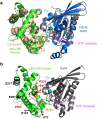
Modeled interface region between the human p85α BH domain and Rab5. (a) Overlay of the crystal structure of human Cdc42GAP (tan) – Cdc42 (blue) complex (PDB ID: 2NGR) with the human p85α BH domain (green; PDB ID: 1PBW) and the human Rab5-GTP analogue (15–184; grey; PDB ID: 1R2Q). The magnesium (teal) and GTP analogue (magenta) bound to Rab5 are indicated. Catalytically important arginine residues within Cdc42GAP (yellow) and p85α BH domain (orange) are shown. (b) Modeled human p85α BH domain (green) – Rab5 (grey) complex. The magnesium (teal) and GTP analogue (magenta) bound to Rab5 are indicated. Key p85α BH domain residues are shown: R151 (orange, important for GAP activity), and residues in red are important for Rab5 binding (L191, V263, R274). The location of patient-derived p85α BH mutations with significant impacts on Rab5 binding are shown in black (E137, I177, E217, E297).

Similar articles

Cited by

References

    1. Yuan TL, Cantley LC. PI3K pathway alterations in cancer: variations on a theme. Oncogene. 2008;27:5497–5510. doi: 10.1038/onc.2008.245. - DOI - PMC - PubMed
    1. Yu J, et al. Regulation of the p85/p110 phosphatidylinositol 3′-kinase: stabilization and inhibition of the p110alpha catalytic subunit by the p85 regulatory subunit. Mol Cell Biol. 1998;18:1379–1387. doi: 10.1128/MCB.18.3.1379. - DOI - PMC - PubMed
    1. Cuevas BD, et al. Tyrosine phosphorylation of p85 relieves its inhibitory activity on phosphatidylinositol 3-kinase. J Biol Chem. 2001;276:27455–27461. doi: 10.1074/jbc.M100556200. - DOI - PubMed
    1. Miled N, et al. Mechanism of two classes of cancer mutations in the phosphoinositide 3-kinase catalytic subunit. Science. 2007;317:239–242. doi: 10.1126/science.1135394. - DOI - PubMed
    1. Cantley LC. The phosphoinositide 3-kinase pathway. Science. 2002;296:1655–1657. doi: 10.1126/science.296.5573.1655. - DOI - PubMed

Publication types

Substances