

Interleukin 17 enhances bone morphogenetic protein-2-induced ectopic bone formation
- PMID: 29740080
- PMCID: PMC5940874
- DOI: 10.1038/s41598-018-25564-9
Interleukin 17 enhances bone morphogenetic protein-2-induced ectopic bone formation
Abstract
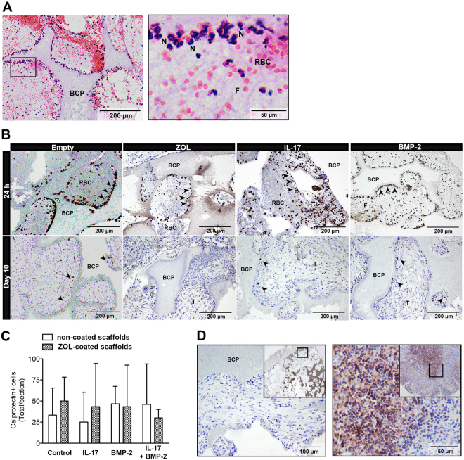
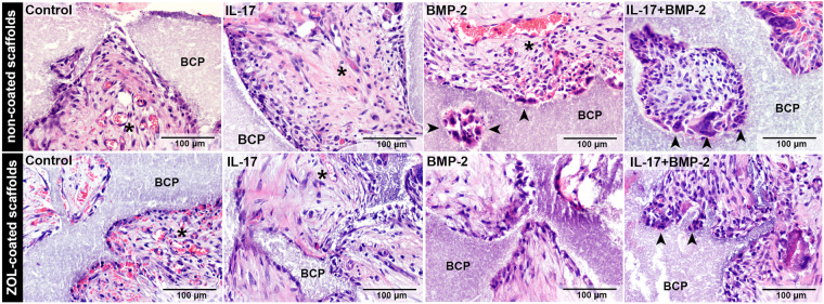
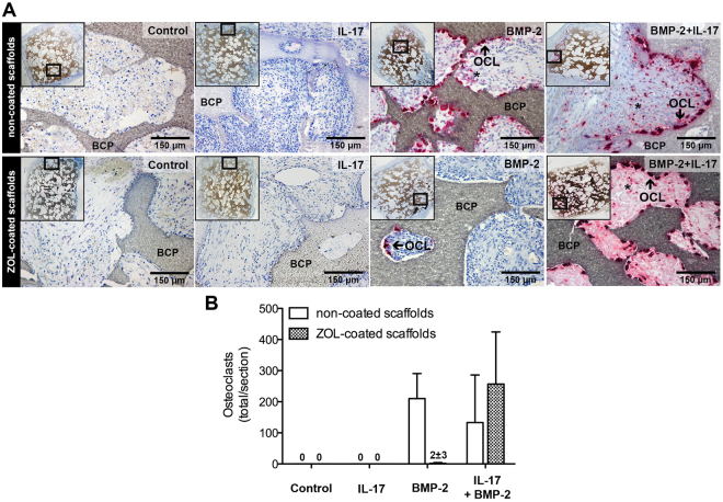
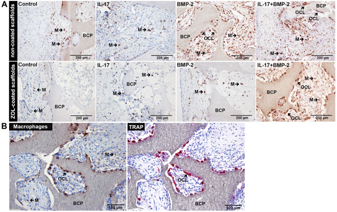
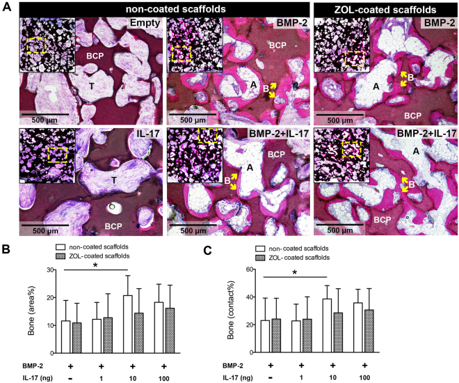
Interleukin 17 (IL-17) stimulates the osteogenic differentiation of progenitor cells in vitro through a synergy with bone morphogenetic protein (BMP)-2. This study investigates whether the diverse responses mediated by IL-17 in vivo also lead to enhanced BMP-2-induced bone formation. Since IL-17 is known to induce osteoclastogenesis, we studied the interactions between IL-17 and BMP-2 in ceramic scaffolds either or not carrying a coating with the bisphosphonate zoledronic acid (ZOL). Histological evaluation revealed that IL-17 alone did not induce any osteoclasts at day 10. On the other hand, BMP-2 clearly stimulated early tissue ingrowth and osteoclastogenesis. Both of these processes were blocked in presence of ZOL. IL-17 signaling restored early vascularized connective tissue formation and osteoclastogenesis induced by BMP-2 in ZOL-coated scaffolds. After 12 weeks, the bone volume induced by co-delivery of BMP-2 and IL-17 was doubled as compared to that induced by BMP-2 alone. We conclude that IL-17 has osteo-stimulatory effects through a synergy with bone-inductive BMP-2. Although local and single application of IL-17 does not mediate osteoclast formation, it could promote other processes involved in bone formation such as connective tissue ingrowth. The use of IL-17 may contribute to the development of improved bone graft substitutes.
Conflict of interest statement
The authors declare no competing interests.
Figures


Similar articles
-
Synergy between IL-6 and soluble IL-6 receptor enhances bone morphogenetic protein-2/absorbable collagen sponge-induced bone regeneration via regulation of BMPRIA distribution and degradation.Biomaterials. 2015 Oct;67:308-22. doi: 10.1016/j.biomaterials.2015.07.047. Epub 2015 Jul 26. Biomaterials. 2015. PMID: 26232880
-
Local co-application of zoledronate promotes long-term maintenance of newly formed bone induced by recombinant human bone morphogenetic protein 2.Biochem Biophys Res Commun. 2016 Nov 18;480(3):314-320. doi: 10.1016/j.bbrc.2016.10.034. Epub 2016 Oct 14. Biochem Biophys Res Commun. 2016. PMID: 27746180
-
Bone morphogenetic protein-2 enhances osteoclast formation mediated by interleukin-1alpha through upregulation of osteoclast differentiation factor and cyclooxygenase-2.Biochem Biophys Res Commun. 1999 May 27;259(1):97-102. doi: 10.1006/bbrc.1999.0715. Biochem Biophys Res Commun. 1999. PMID: 10334922
-
Identification of cell cycle-arrested quiescent osteoclast precursors in vivo.Adv Exp Med Biol. 2010;658:21-30. doi: 10.1007/978-1-4419-1050-9_3. Adv Exp Med Biol. 2010. PMID: 19950012 Review.
-
Factors that modulate the effects of bone morphogenetic protein-induced periodontal regeneration: a critical review.J Periodontol. 2002 Aug;73(8):925-36. doi: 10.1902/jop.2002.73.8.925. J Periodontol. 2002. PMID: 12211503 Review.
Cited by
-
Use of Therapeutic Pathogen Recognition Receptor Ligands for Osteo-Immunomodulation.Materials (Basel). 2021 Feb 27;14(5):1119. doi: 10.3390/ma14051119. Materials (Basel). 2021. PMID: 33673651 Free PMC article.
-
Regulation of Osteoblast Differentiation by Cytokine Networks.Int J Mol Sci. 2021 Mar 11;22(6):2851. doi: 10.3390/ijms22062851. Int J Mol Sci. 2021. PMID: 33799644 Free PMC article. Review.
-
Regulation of the mesenchymal stem cell fate by interleukin-17: Implications in osteogenic differentiation.World J Stem Cells. 2021 Nov 26;13(11):1696-1713. doi: 10.4252/wjsc.v13.i11.1696. World J Stem Cells. 2021. PMID: 34909118 Free PMC article. Review.
-
Elevated BMPR2 expression amplifies osteoblast differentiation in ankylosing spondylitis.J Rheum Dis. 2023 Oct 1;30(4):243-250. doi: 10.4078/jrd.2023.0024. Epub 2023 Jul 28. J Rheum Dis. 2023. PMID: 37736586 Free PMC article.
-
The Interrelationship Between Diabetes, IL-17 and Bone Loss.Curr Osteoporos Rep. 2020 Feb;18(1):23-31. doi: 10.1007/s11914-020-00559-6. Curr Osteoporos Rep. 2020. PMID: 32002770 Free PMC article. Review.
References
-
- Govender S, et al. Recombinant human bone morphogenetic protein-2 for treatment of open tibial fractures: a prospective, controlled, randomized study of four hundred and fifty patients. The Journal of bone and joint surgery American volume. 2002;84-A:2123–2134. doi: 10.2106/00004623-200212000-00001. - DOI - PubMed
Publication types
MeSH terms
Substances
LinkOut - more resources
Full Text Sources
Other Literature Sources
