Review
. 2018 Jun;19(6):351-367.
doi: 10.1038/s41583-018-0003-6.

Breathing matters

Affiliations
Review

Breathing matters

Christopher A Del Negro et al. Nat Rev Neurosci. 2018 Jun.

Abstract

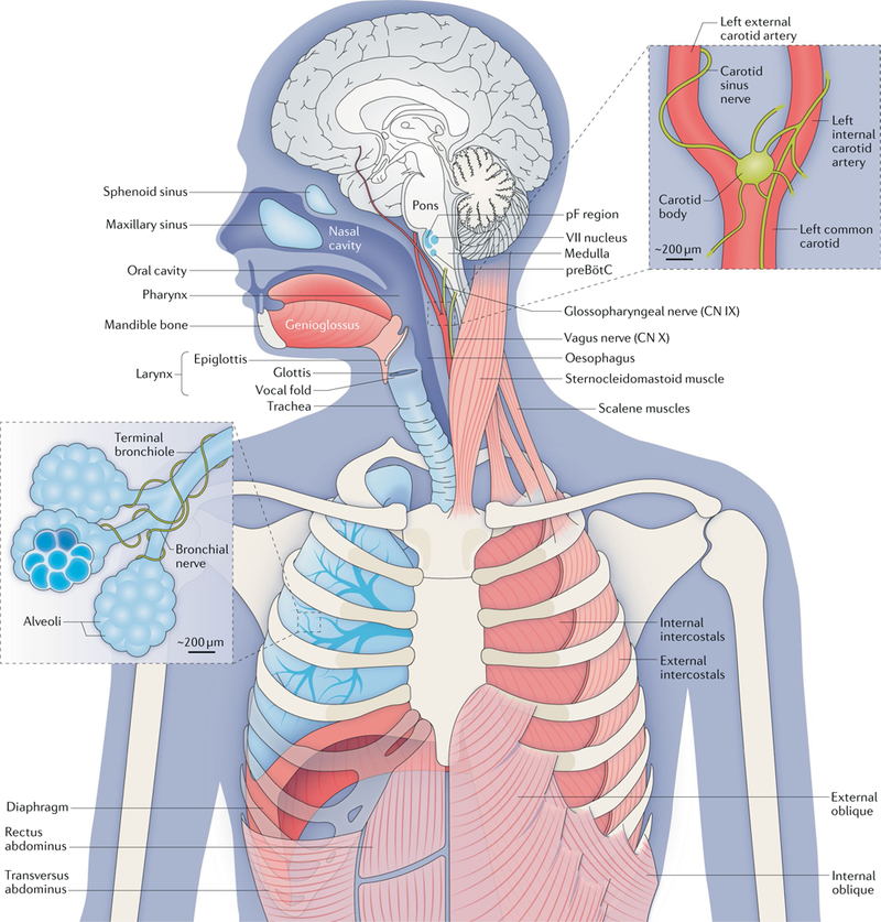
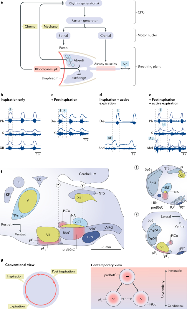
Breathing is a well-described, vital and surprisingly complex behaviour, with behavioural and physiological outputs that are easy to directly measure. Key neural elements for generating breathing pattern are distinct, compact and form a network amenable to detailed interrogation, promising the imminent discovery of molecular, cellular, synaptic and network mechanisms that give rise to the behaviour. Coupled oscillatory microcircuits make up the rhythmic core of the breathing network. Primary among these is the preBötzinger Complex (preBötC), which is composed of excitatory rhythmogenic interneurons and excitatory and inhibitory pattern-forming interneurons that together produce the essential periodic drive for inspiration. The preBötC coordinates all phases of the breathing cycle, coordinates breathing with orofacial behaviours and strongly influences, and is influenced by, emotion and cognition. Here, we review progress towards cracking the inner workings of this vital core.


Conflict of interest statement

Competing interests

The authors declare no competing interests.

Figures

Fig. 1 |
Fig. 1 |. The anatomy and physiology of respiration.
Breathing movements depend on pump, resistance and accessory muscles. Pump muscles include the dome-like, uniquely mammalian diaphragm and the external intercostals. During inspiration, descent of the diaphragm combined with rib elevation by the external intercostals expands the lungs to draw in air (assuming the airways are patent). The oblique abdominals and transversus abdominus and internal intercostals are expiratory pump muscles. Airway resistance muscles, which modulate inspiratory and expiratory airflow, include skeletal muscles of the tongue (including the genioglossus (shown) and the hyoglossus, styloglossus and stylohyoid muscles (not shown)), glottis, larynx and pharynx, as well as smooth muscle of the bronchi. The sternocleidomastoid and scalene muscles are accessory muscles that stabilize the rib cage. The gas-exchanging surface of human lungs, consisting of ~5 × 10⁸ alveoli each measuring 200 μm in diameter, is roughly half the size of a tennis court (~70 m2) but is contained in a volume of <3 litres. At rest, we inhale and exhale ~5 litres of air per minute (~10 × 500 millilitre breaths per minute, containing ~ 1 litre of O2); we extract from the inspired air ~250 millilitres of O2 per minute to support metabolism and add to the expired air ~200 millilitres of CO2 per minute. The regulation of breathing relies on feedback from peripheral and central chemosensors. Carotid bodies, at the branch point of the carotid arteries, monitor the partial pressure of O2 (pO2), the partial pressure of CO2 (pCO2) and pH in arterial blood and signal to the brainstem via the glossopharyngeal nerve (cranial nerve (CN) IX). In the brainstem, chemosensory neurons and glia in the ventral parafacial nucleus (pFV) and other regions detect and respond to fluctuations in CO2 levels and pH in the cerebrospinal fluid. These neurons project paucisynaptically to the preBötzinger Complex (preBötC) and other sites to influence breathing to maintain homeostasis. Breathing in mammals (and other air breathers) under most conditions is extremely sensitive to changes in CO2 levels that directly affect pH. For example, in resting humans, an ~2.5% increase in pCO2 from 40 to 41 mmHg will increase ventilation ~40% from ~5 to ~7 litres per minute. By contrast, breathing is relatively insensitive to changes in O2 levels at rest. The O2–haemog1obin dissociation curve is fairly flat at normal levels of arterial O2 (100 ± 20 mm Hg), meaning that haemoglobin is >96% saturated with O2 even if breathing increases substantially. However, under certain conditions (for example, high altitude or intense exercise), hypoxia provides a powerful stimulus to breathe. Continuous breathing comes at a considerable metabolic cost insofar as the respiratory muscles are the only skeletal muscles that are active during all sleep and wake states. To counter this, mammals evolved a powerful diaphragm that is sufficient to inflate the lungs, and a resting breathing pattern in which inspiration (and usually postinspiration) is active (requiring muscle contraction), whereas expiration is passive. In birds (and most lower vertebrates) that do not have a diaphragm, inspiration and expiration are both active at rest. Central breathing networks are also modulated by mechanosensory feedback regarding the status of the pump muscles and lungs. Stretch- receptor afferents in airway smooth muscle encode volume-related information via the vagus nerve (CN X), which is crucial in maintaining optimal lung volumes for efficient breathing. This feedback also underlies Breuer-Hering reflexes, which are important in controlling the timing and pattern of each breath. Lung stretch-receptor afferents and their central relay interneurons rhythmically inhibit the inspiratory preBötC and excite the expiratory lateral parafacial nucleus (pFL) when lungs are inflated (inspiratory termination reflex) and conversely excite the preBötC and inhibit the pFL when lungs are deflated. VII nucleus, facial motor nucleus. The figure is based on a drawing contributed by J. Milstein.
Fig. 2 |
Fig. 2 |. Elements of the breathing central pattern generator. a
a | Schematic view of breathing neural and physical plant. The image shows a central pattern generator (CPG) composed of rhythm-generating and pattern-generating microcircuits. Spinal and cranial motor nuclei innervate pump and airway muscles, respectively. Airways, lungs, blood gases and sensory (‘chemo’ and ‘mechano’) feedback are indicated. b | Inspiration (I) is the inexorable phase of the breathing cycle. In an anaesthetized rat, the inspiratory cycle is evident in electrical recordings (raw and integrated signals) from the phrenic nerve (Ph), a branch of the vagus nerve (cranial nerve (CN) X) innervating laryngeal adductor muscles, and the hypoglossal nerve (CN XII) innervating the genioglossus muscle (tongue protruder). c | Also in an anesthetized rat, inspiration recorded via diaphragmatic (Dia) electromyography (EMG), shown with postinspiration (PI) recorded from the laryngeal branch of CN X. d | Also in an anesthetized rat, inspiration shown via diaphragmatic EMG (Dia), with active expiration (AE; evoked by disinhibition of the lateral parafacial respiratory group (pFL)) indicated by abdominal EMG (Abd). e | In a reduced in situ rat preparation, inspiration, postinspiration and active expiration measured via electrical recordings from the phrenic (Ph) nerve, CN X and lumbar abdominal (Abd) nerve, respectively. Active expiration was evoked by hypercapnia. f | Parasagittal view of the brainstem containing the breathing CPG. Respiratory rhythmogenic sites are shown in red: the preBötzinger Complex (preBötC; inspiratory), the pFL (expiratory) and the more medial chemosensitive ventral parafacial respiratory group (pFV; rhythmogenic in the perinatal period only), as well as the ‘postinspiratory comp1ex’(PiCo; hypothesized to underlie postinspiration). Other rhythmogenic sites, such as the whisking-related ventral intermediate reticular formation (vIRT) and the masticatory trigeminal principal sensory nucleus (NVsnpr), are shown in blue. Cranial motor nuclei controlling airway resistance muscles, the hypoglossal motor nucleus (XII) and the nucleus ambiguus (NA), as well as facial muscles, the facial motor nucleus (VII) and the trigeminal motor nucleus (V), are shown in green. Brainstem sites associated with breathing motor pattern or sensorimotor integration are shown in grey: the rostral ventral respiratory group (rVRG) containing inspiratory, that is, phrenic and external intercostal, premotor neurons and the caudal ventral respiratory group (rVRG) containing expiratory premotor neurons. Other sites include the pontine Kölliker-Fuse nucleus (KF) and parabrachial nucleus (PB), the nucleus of the solitary tract (NTS) and the expiratory BotC. Also shown is the locus coeruleus (LC) that receives preBötC projections, the cerebellum and the lateral reticular nucleus (LRN). Insets 1 and 2 show transverse sections at the level of the preBötC (dotted line 1) and pF (dotted line 2). Additional structures in the insets include the spinal trigeminal tract (Sp5), the spinal trigeminal sensory nucleus oralis (Sp5O) and the interpolaris (Sp5I), the inferior olive (IO) and the pyramidal tract (pyr). g | In the conventional view of breathing, inspiration, postinspiration and expiration constitute a continuous unitary breathing cycle. According to the contemporary view, inspiration is the inexorable part of the breathing cycle, whereas postinspiration and expiration are conditional, driven by three distinct, coupled oscillators. When all parts of the cycle are manifest, the preBötC coordinates the phases. Part a is adapted with permission from REF, Elsevier. Part b is adapted with permission from APS, REF. Part c is republished with permission of Society for Neuroscience, from Role of inhibition in respiratory pattern generation, Janczewski, W. A. et al. 33 (13), 2013 (REF); permission conveyed through Copyright Clearance Center, Inc. Part d is republished with permission of Society for Neuroscience, from Active expiration induced by excitation of ventral medulla in adult anesthetized rats, Pagliardini, S. et al. 31 (8), 2011 (REF.); permission conveyed through Copyright Clearance Center, Inc. Part e is adapted with permission from REF., Elsevier.
Fig. 3 |
Fig. 3 |. Emergent network rhythms and burstlet theory.
a | In vitro recordings of preBötzinger Complex (preBötC) field potentials and hypoglossal nerve (cranial nerve XII) activity showing rhythmic inspiratory-related output at several levels of excitability determined by changes in the concentration of K+ in the bathing solution. Bursts are associated with full- amplitude activity synchronized in the preBötC and XII nerve root. Lower-amplitude burstlets (without XII output) are marked with red triangles. b | Schematic of network activity underlying burstlets, bursts and XII output. Numerals indicate different stages of the trajectories in part a: 1, refractory state following inspiration; 2, spontaneous spiking resumes in some neuronal constituents; 3, active neurons are mutually reinforcing owing to recurrent synaptic interconnections, resulting in low-amplitude pre-inspiratory activity that percolates through the rhythmogenic population (that is, a burstlet); and 4, the burstlet triggers a full burst, which propagates to pattern-related preBötC neurons, premotor neurons and motor neurons, generating XII motor output. Colour scale: blue reflects quiescence, and pink and red map to progressively higher rates of spiking. Lines connecting the cells represent strength of synaptic drive: the absence of a line reflects synaptic depression; dotted lines reflect light synaptic drive; and solid lines represent strong drive. c | The interburst interval (IBI) in part a (shown at 9 mM K+) is reproduced here to illustrate the role of spike synchronization, schematized by three neurons projecting to a fourth neuron. Left: when action potentials (APs) are unsynchronized, excitatory postsynaptic potentials (EPSPs) do not summate. Middle and right: as the constituent neurons fire APs in progressively greater synchrony, the summation of EPSPs progressively increases, generating more spikes in postsynaptic partners. We suggest that greater synchrony among preBötC neurons ensues during the IBI. Part a is republished with permission of Society of Neuroscience, from Distinct inspiratory rhythm and pattern generating mechanisms in the preBötzinger complex, Kam, K. et al. 33 (22), 2013 (REF); permission conveyed through Copyright Clearance Center, Inc. Part c is adapted from REF, by permission of Oxford University Press.
Fig. 4 |
Fig. 4 |. A circuit that generates and modulates sighs.
a | Gastrin-releasing peptide (GRP) and neuromedin B (NMB)-expressing neurons of the parafacial nucleus (pF) project to preBötzinger Complex (preBötC) neurons that express GRP receptors (GRPRs), NMB receptors (NMBRs) or both. b | Breathing patterns in an anaesthetized adult rat showing tidal volume (VT; maximal excursion 5 ml), annotated to differentiate an eupnoeic breath from a sigh, and diaphragmatic electromyography (Dia trace shows raw recording; ∫Dia trace shows integrated activity, in arbitrary units). c | Time- compressed recordings of VT and ∫ Dia. Thick baseline reflects continuous eupnoea. Large periodic spikes are sighs, which increase in frequency following infusion of NMB. LRN, lateral reticular nucleus; NA, nucleus ambiguus; pFV, ventral parafacial nucleus; PiCo, postinspiratory complex; rVRG, rostral ventral respiratory group; VII, facial motor nucleus; vIRT, ventral intermediate reticular formation. Part a and c are adapted from ref. , Macmillan Publishers Limited. Part b is adapted with permission from ref. , Elsevier.

References

    1. Smith JC, Ellenberger HH, Ballanyi K, Richter DW & Feldman JL Pre-Bötzinger complex: a brainstem region that may generate respiratory rhythm in mammals. Science 254, 726–729 (1991). This seminal work announced the preBötC, which was identified and named in reference 15 cited therein. - PMC - PubMed
    1. Schwarzacher SW, Smith JC & Richter DW Pre-Bötzinger complex in the cat. J. Neurophysiol. 73, 1452–1461 (1995). - PubMed
    1. Schwarzacher SW, Rüb U & Deller T Neuroanatomical characteristics of the human pre-Bötzinger Complex and its involvement in neurodegenerative brainstem diseases. Brain J. Neurol. 134, 24–35 (2011). - PubMed
    1. Tupal S et al. Testing the role of preBötzinger complex somatostatin neurons in respiratory and vocal behaviors. Eur. J. Neurosci. 40, 3067–3077 (2014). - PMC - PubMed
    1. Wenninger JM et al. Large lesions in the pre- Bötzinger complex area eliminate eupneic respiratory rhythm in awake goats. J. Appl. Physiol. 97, 1629–1636 (2004). - PubMed