. 2018 Apr 24:9:755.
doi: 10.3389/fmicb.2018.00755. eCollection 2018.

The Membrane-Bound C Subunit of Reductive Dehalogenases: Topology Analysis and Reconstitution of the FMN-Binding Domain of PceC

Affiliations

The Membrane-Bound C Subunit of Reductive Dehalogenases: Topology Analysis and Reconstitution of the FMN-Binding Domain of PceC

Géraldine F Buttet et al. Front Microbiol. .

Abstract

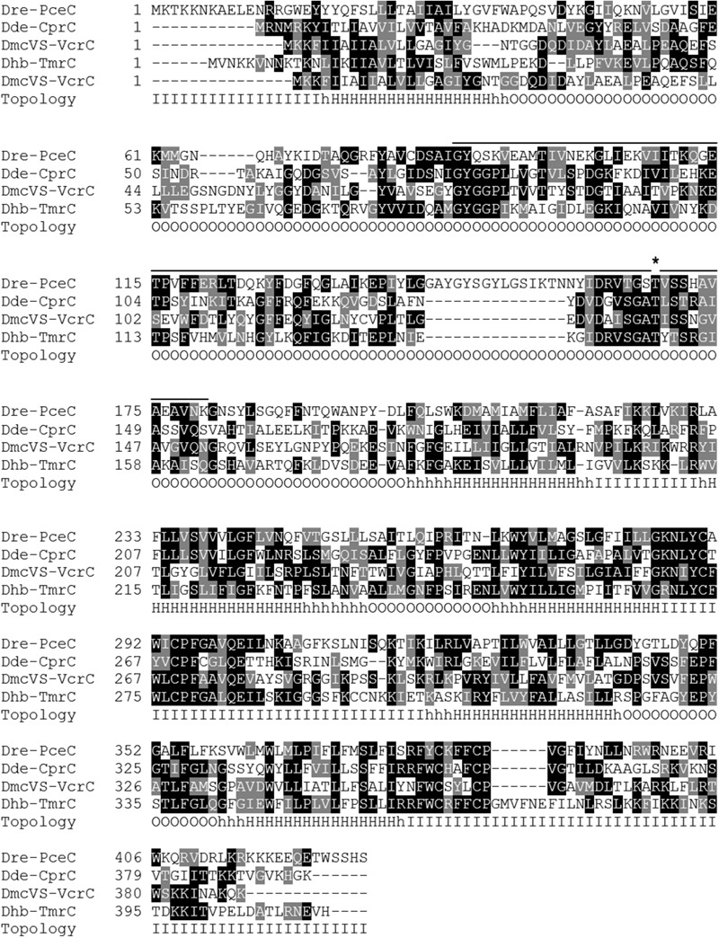
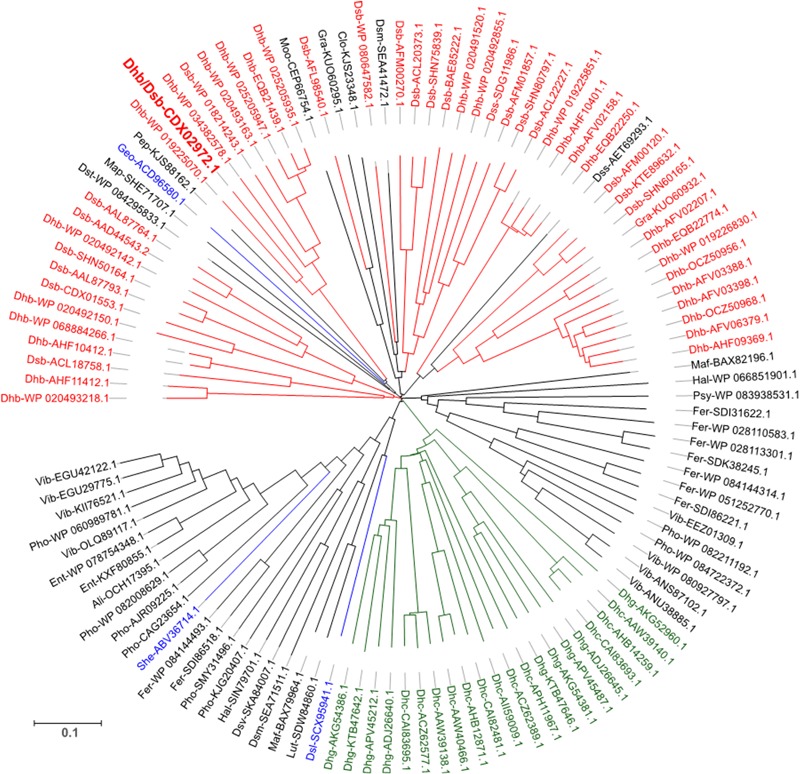
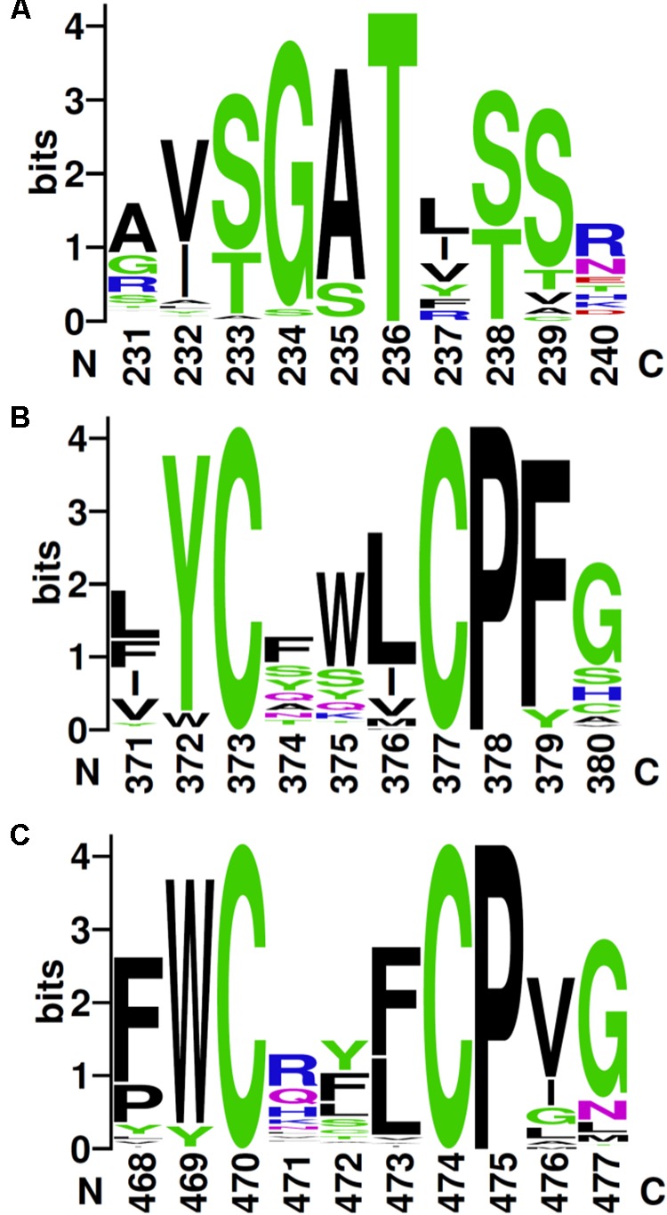
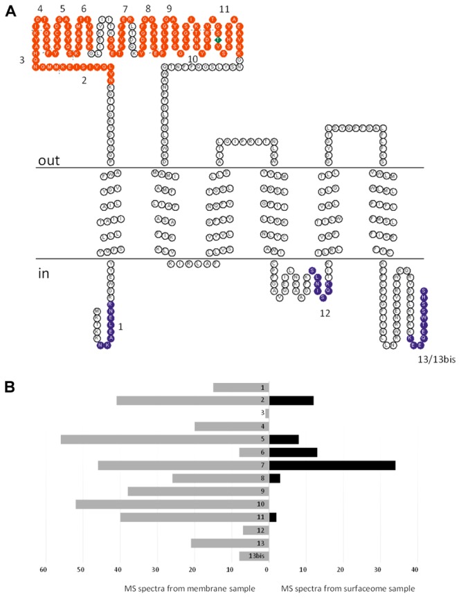
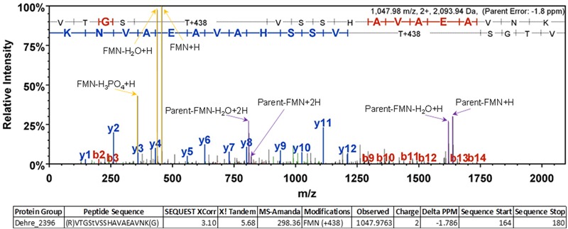
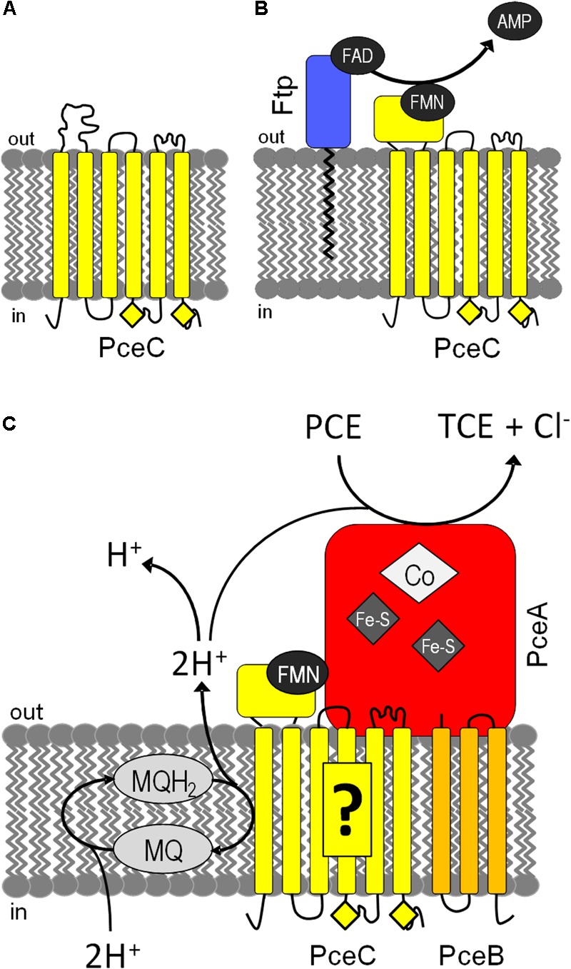
Organohalide respiration (OHR) is the energy metabolism of anaerobic bacteria able to use halogenated organic compounds as terminal electron acceptors. While the terminal enzymes in OHR, so-called reductive dehalogenases, are well-characterized, the identity of proteins potentially involved in electron transfer to the terminal enzymes remains elusive. Among the accessory genes identified in OHR gene clusters, the C subunit (rdhC) could well code for the missing redox protein between the quinol pool and the reductive dehalogenase, although it was initially proposed to act as transcriptional regulator. RdhC sequences are characterized by the presence of multiple transmembrane segments, a flavin mononucleotide (FMN) binding motif and two conserved CX3CP motifs. Based on these features, we propose a curated selection of RdhC proteins identified in general sequence databases. Beside the Firmicutes from which RdhC sequences were initially identified, the identified sequences belong to three additional phyla, the Chloroflexi, the Proteobacteria, and the Bacteriodetes. The diversity of RdhC sequences mostly respects the phylogenetic distribution, suggesting that rdhC genes emerged relatively early in the evolution of the OHR metabolism. PceC, the C subunit of the tetrachloroethene (PCE) reductive dehalogenase is encoded by the conserved pceABCT gene cluster identified in Dehalobacter restrictus PER-K23 and in several strains of Desulfitobacterium hafniense. Surfaceome analysis of D. restrictus cells confirmed the predicted topology of the FMN-binding domain (FBD) of PceC that is the exocytoplasmic face of the membrane. Starting from inclusion bodies of a recombinant FBD protein, strategies for successful assembly of the FMN cofactor and refolding were achieved with the use of the flavin-trafficking protein from D. hafniense TCE1. Mass spectrometry analysis and site-directed mutagenesis of rFBD revealed that threonine-168 of PceC is binding FMN covalently. Our results suggest that PceC, and more generally RdhC proteins, may play a role in electron transfer in the metabolism of OHR.

Keywords: PceC; RdhC; flavin mononucleotide (FMN); flavin transferase; flavin-trafficking proteins (Ftp); flavoproteins; organohalide respiration; protein reconstitution.


Figures

FIGURE 1
FIGURE 1
Sequence alignment of four typical members of the RdhC enzyme family: PceC (CAG70347.1) of Dehalobacter restrictus (Dre); CprC (AAD44543.2) of Desulfitobacterium dehalogenans (Dde); VcrC (ACZ62389.1) of Dehalococcoides mccartyi strain VS (DmcVS); TmrC (WP_021315090.1) of Dehalobacter sp. strain UNSWDHB (Dhb). The predicted topology is indicated under the alignment: O, outside; I, inside; H, transmembrane α-helices. The bold line above the alignment indicates the predicted FMN-binding domain (FBD, smart00900) with the conserved threonine residue predicted to bind FMN covalently (indicated by the asterisk).
FIGURE 2
FIGURE 2
Sequence likelihood analysis of 117 representative RdhC proteins. Sequences are annotated with an abbreviation of the corresponding species followed by the reference number. The sequence annotated in bold red is PceC from Desulfitobacterium hafniense, the representative sequence for the protein under investigation in this study. Colors indicate sequences belonging to well-described OHR bacterial genera: red, Dehalobacter and Desulfitobacterium; green, Dehalococcoides and Dehalogenimonas; blue, additional OHRB. Detailed information on the overall database used is available in Supplementary Material (Supplementary Table 2).
FIGURE 3
FIGURE 3
Conserved sequence motifs in RdhC family. Based on the alignment of the 117 RdhC non-redundant representative sequences, motifs were drawn using Weblogo: (A) the FMN-binding motif, with the fully conserved threonine involved in binding FMN covalently; (B,C) the two CX3P signatures. Residue numbering follows the alignment.
FIGURE 4
FIGURE 4
Topology analysis of D. restrictus PceC from shaving experiment. (A) The predicted topology of PceC is shown. In red are indicated the peptides detected at the surface of D. restrictus cells, while in purple the ones exclusively detected in the membrane sample. (B) Graphical view of the number of detected peptides in the membrane sample (in gray) and in the surfaceome (in black).
FIGURE 5
FIGURE 5
Representative MS/MS spectrum confirming the FMN localization at position T168 of PceC. Diagnostic ions corresponding to the FMN+H and its fragments are highlighted in yellow allowing unambiguous identification of the post-translational modification.
FIGURE 6
FIGURE 6
Reconstitution strategies for rFBD. (A) Reconstitution of rFBD by reverse urea gradient on Ni-NTA column. For the reconstitution steps, only the first (R1) and the last sample (R10) are shown. Legend: L, protein ladder; R#, 10 successive gradient steps with decreasing urea concentration; W, wash step; E#, successive eluted samples; U, sample eluted with 4 M urea. (B) Reconstitution of rFBD by dialyzing out urea in a stepwise manner. For both (A,B), the top panels show Coomassie blue stained gels, while the bottom panels display the same gels under UV illumination. Stars indicate rFBD proteins.
FIGURE 7
FIGURE 7
Reconstitution of rFBD wild-type (WT) and threonine-168 variant (T168V). The top panels show Coomassie blue stained gels, while the bottom panels display the same gels under UV illumination. Stars indicate rFBD proteins. L, protein ladder.
FIGURE 8
FIGURE 8
Tentative models for the assembly and function of PceC in D. hafniense. (A) PceC (in yellow) is likely inserted in the cytoplasmic membrane while the FBD is in an unfolded state. (B) On the exocytoplasmic face of the membrane, Ftp (in blue) catalyzes the hydrolysis of FAD to FMN and AMP, and attaches FMN to the threonine-168 of PceC, resulting in the folding of the FBD. (C) PceC is hypothesized to build a protein complex with PceA (in red) and PceB (in orange), and to play the role of electron transfer from the menaquinol (MQ) pool to the redox centers of PceA via the FMN cofactor. Yellow diamonds represent the conserved CX3CP sequence motifs.

References

    1. Adrian L., Löffler F. (2016). Organohalide-Respiring Bacteria. Berlin: Springer-Verlag.
    1. Backiel J., Juarez O., Zagorevski D. V., Wang Z., Nilges M. J., Barquera B. (2008). Covalent binding of flavins to RnfG and RnfD in the Rnf complex from Vibrio cholerae. Biochemistry 47 11273–11284. 10.1021/bi800920j - DOI - PMC - PubMed
    1. Beck B. J., Downs D. M. (1998). The apbE gene encodes a lipoprotein involved in thiamine synthesis in Salmonella typhimurium. J. Bacteriol. 180 885–891. - PMC - PubMed
    1. Bertsova Y. V., Fadeeva M. S., Kostyrko V. A., Serebryakova M. V., Baykov A. A., Bogachev A. V. (2013). Alternative pyrimidine biosynthesis protein ApbE is a flavin transferase catalyzing covalent attachment of FMN to a threonine residue in bacterial flavoproteins. J. Biol. Chem. 288 14276–14286. 10.1074/jbc.M113.455402 - DOI - PMC - PubMed
    1. Borrero-de Acuna J. M., Timmis K. N., Jahn M., Jahn D. (2017). Protein complex formation during denitrification by Pseudomonas aeruginosa. Microb. Biotechnol. 10 1523–1534. 10.1111/1751-7915.12851 - DOI - PMC - PubMed

LinkOut - more resources