. 2018 May 8;4(1):45.
doi: 10.1186/s40792-018-0454-z.

A case of mixed adenoneuroendocrine carcinoma (MANEC) arising in Barrett's esophagus: literature and review

Affiliations

A case of mixed adenoneuroendocrine carcinoma (MANEC) arising in Barrett's esophagus: literature and review

Tetsuro Kawazoe et al. Surg Case Rep. .

Abstract

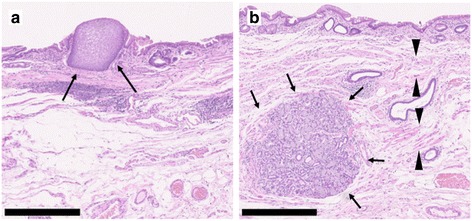
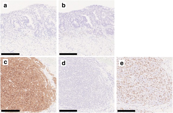
Background: Mixed adenoneuroendocrine carcinoma (MANEC) is defined as a neoplasm composed of both exocrine and endocrine carcinomas, each comprising at least 30% of the tumor. MANEC can occur in various organs of the gastrointestinal tract, including the esophagus, stomach, and colon. We herein provide the first case report of surgically resected MANEC arising in Barrett's esophagus (BE).

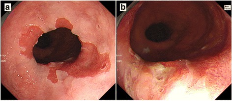
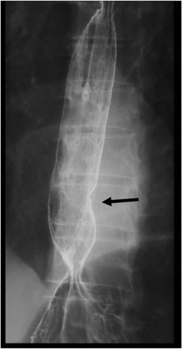
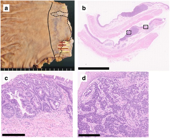
Case presentation: A 70-year-old man presenting with abdominal pain was referred to our hospital. Upper endoscopy showed a type 0-IIa + IIc elevated lesion adjacent to BE. According to a biopsy specimen, the elevated lesion was diagnosed as adenocarcinoma with neuroendocrine differentiation. No lymphatic or distant metastasis was detected in the preoperative examination. Laparoscopic distal esophagectomy and proximal gastrectomy were performed, and a diagnosis of MANEC in BE was determined according to the surgically resected specimen.

Conclusions: A very rare case of MANEC in BE was detected. BE can be the origin of esophageal MANEC as well as adenocarcinoma. Due to the small number of esophageal or esophagogastric MANEC cases reported, further accumulation of such cases is necessary to recommend an optimal management strategy for esophageal or esophagogastric MANEC.

Keywords: Barrett’s esophagus; Mixed adenoneuroendocrine carcinoma (MANEC); Neuroendocrine tumor.


Conflict of interest statement

Ethics approval and consent to participate

The present study was conducted in accordance with the ethical standards of our institution.

Consent for publication

Written informed consent was obtained from the patient for publication of this case report and any accompanying images.

Competing interests

The authors declare that they have no competing interests.

Publisher’s Note

Springer Nature remains neutral with regard to jurisdictional claims in published maps and institutional affiliations.

Figures

Fig. 1
Fig. 1
Upper endoscopy. Upper endoscopy showed an elongated columnar epithelium from the squamocolumnar junction indicating BE (a) and a type 0-IIa + IIc elevated lesion adjacent to the BE lesion (b)
Fig. 2
Fig. 2
Esophagography. Esophagography showed a type 0-IIa + IIc elevated lesion (15 mm in size) on the left wall of the lower esophagus, and the tumor exhibited arcuate change suggesting submucosal invasion
Fig. 3
Fig. 3
Histological findings of MANEC. Macroscopically, a type 0-IIa + IIc lesion measuring 25 × 10 mm was detected in the esophagogastric junction. The black solid line indicates the esophagogastric junction, the black dotted line the squamocolumnar junction, the yellow line the adenocarcinoma component, and the red line the NEC component (a). A loupe image of the lesion is shown (b, scale bar 10 nm). The solid rectangle indicates well differentiated adenocarcinoma (c scale bar 250 μm), and the dotted rectangle indicates an area of proliferation of round-shaped carcinoma cells with hyperchromatic nuclei and scant cytoplasm in a nested pattern, indicative of small cell NEC (d scale bar 250 μm)
Fig. 4
Fig. 4
Histological findings of Barrett’s esophagus. BE was recognized histologically. Some islands of squamous epithelium (a, arrow), esophageal glands beneath the columnar epithelium (b, arrow), and double-layered muscularis mucosae (b, arrow head) were observed. All scale bars—500 μm
Fig. 5
Fig. 5
Immunohistochemical findings. Immunohistochemically, adenocarcinoma cells were negative for synaptophysin (a) and chromogranin A (b), while the round-shaped carcinoma cells were diffusely positive for synaptophysin (c), but negative for chromogranin A (d). The Ki67 labeling index was 50% (e). All scale bars—250 μm

References

    1. Kunz PL. Carcinoid and neuroendocrine tumors: building on success. J Clin Oncol. 2015;33(16):1855–1863. doi: 10.1200/JCO.2014.60.2532. - DOI - PubMed
    1. Bosman FT, Carneiro F, Hruban RH, Theise ND. WHO classification of tumours of the digestive system. 4th ed. Geneva: World Health Organization; 2010. https://www.cabdirect.org/cabdirect/abstract/20113051318.
    1. Kadhim MM, Jespersen ML, Pilegaard HK, Nordsmark M, Villadsen GE. Mixed adenoneuroendocrine carcinoma is a rare but important tumour found in the oesophagus. Case Rep Gastrointest Med. 2016;2016:9542687. - PMC - PubMed
    1. Yamauchi H, Sakurai S, Nakazawa N, Yoshida T, Tabe Y, Saitoh K, et al. A case of mixed adenoneuroendocrine carcinoma of the stomach with focal intestinal metaplasia and hypergastrinemia. Int Surg. 2015;100(3):562–567. doi: 10.9738/INTSURG-D-14-00074.1. - DOI - PMC - PubMed
    1. Kwok CM. Mixed adenoneuroendocrine carcinoma of the stomach. Case Rep Gastroenterol. 2015;9(2):241–245. doi: 10.1159/000437293. - DOI - PMC - PubMed

LinkOut - more resources