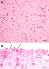
Alexander disease: an astrocytopathy that produces a leukodystrophy
- PMID: 29740945
- PMCID: PMC8028392
- DOI: 10.1111/bpa.12601
Alexander disease: an astrocytopathy that produces a leukodystrophy
Abstract
Alexander Disease (AxD) is a degenerative disorder caused by mutations in the GFAP gene, which encodes the major intermediate filament of astrocytes. As other cells in the CNS do not express GFAP, AxD is a primary astrocyte disease. Astrocytes acquire a large number of pathological features, including changes in morphology, the loss or diminution of a number of critical astrocyte functions and the activation of cell stress and inflammatory pathways. AxD is also characterized by white matter degeneration, a pathology that has led it to be included in the "leukodystrophies." Furthermore, variable degrees of neuronal loss take place. Thus, the astrocyte pathology triggers alterations in other cell types. Here, we will review the neuropathology of AxD and discuss how a disease of astrocytes can lead to severe pathologies in non-astrocytic cells. Our knowledge of the pathophysiology of AxD will also lead to a better understanding of how astrocytes interact with other CNS cells and how astrocytes in the gliosis that accompanies many neurological disorders can damage the function and survival of other cells.
Keywords: Alexander disease; GFAP; astrocytes; leukodystrophy.
© 2018 International Society of Neuropathology.
Figures


Similar articles
-
Disorders of Astrocytes: Alexander Disease as a Model.Annu Rev Pathol. 2017 Jan 24;12:131-152. doi: 10.1146/annurev-pathol-052016-100218. Annu Rev Pathol. 2017. PMID: 28135564 Review.
-
GFAP Mutations in Astrocytes Impair Oligodendrocyte Progenitor Proliferation and Myelination in an hiPSC Model of Alexander Disease.Cell Stem Cell. 2018 Aug 2;23(2):239-251.e6. doi: 10.1016/j.stem.2018.07.009. Cell Stem Cell. 2018. PMID: 30075130 Free PMC article.
-
Astrocyte pathology in Alexander disease causes a marked inflammatory environment.Acta Neuropathol. 2015 Oct;130(4):469-86. doi: 10.1007/s00401-015-1469-1. Epub 2015 Aug 22. Acta Neuropathol. 2015. PMID: 26296699
-
Phenotypic conversions of "protoplasmic" to "reactive" astrocytes in Alexander disease.J Neurosci. 2013 Apr 24;33(17):7439-50. doi: 10.1523/JNEUROSCI.4506-12.2013. J Neurosci. 2013. PMID: 23616550 Free PMC article.
-
Alexander disease: putative mechanisms of an astrocytic encephalopathy.Cell Mol Life Sci. 2004 Feb;61(3):369-85. doi: 10.1007/s00018-003-3143-3. Cell Mol Life Sci. 2004. PMID: 14770299 Free PMC article. Review.
Cited by
-
Long-term survival of a dog with Alexander disease.J Vet Med Sci. 2020 Dec 5;82(11):1704-1707. doi: 10.1292/jvms.20-0133. Epub 2020 Oct 15. J Vet Med Sci. 2020. PMID: 33055453 Free PMC article.
-
The Role of TDP-43 in Neurodegenerative Disease.Mol Neurobiol. 2022 Jul;59(7):4223-4241. doi: 10.1007/s12035-022-02847-x. Epub 2022 May 2. Mol Neurobiol. 2022. PMID: 35499795 Review.
-
A case report of adult-onset Alexander disease clinically presenting as Parkinson's disease: is the comorbidity associated with genetic susceptibility?BMC Neurol. 2020 Jan 17;20(1):27. doi: 10.1186/s12883-020-1616-8. BMC Neurol. 2020. PMID: 31952467 Free PMC article.
-
Lack of astrocytes hinders parenchymal oligodendrocyte precursor cells from reaching a myelinating state in osmolyte-induced demyelination.Acta Neuropathol Commun. 2020 Dec 24;8(1):224. doi: 10.1186/s40478-020-01105-2. Acta Neuropathol Commun. 2020. PMID: 33357244 Free PMC article.
-
Gene and Cellular Therapies for Leukodystrophies.Pharmaceutics. 2023 Oct 24;15(11):2522. doi: 10.3390/pharmaceutics15112522. Pharmaceutics. 2023. PMID: 38004502 Free PMC article. Review.
References
-
- Alexander WS (1949) Progressive fibrinoid degeneration of fibrillary astrocytes associated with mental retardation in a hydrocephalic infant. Brain 72:373–381. - PubMed
-
- Back SA, Tuohy TMF, Chen H, Wallingford N, Craig A, Struve J et al (2005) Hyaluronan accumulates in de‐ myelinated lesions and inhibits oligodendrocyte progenitor maturation. Nat Med 11:966–972. - PubMed
-
- Bellot‐Saez A, Kékesi O, Morley JW, Buskila Y (2017) Astrocytic modulation of neuronal excitability through K+ spatial buffering. Neurosci Biobehav Rev 77:87–97. - PubMed
-
- Borrett D, Becker LE (1985) Alexander's disease. A disease of astrocytes. Brain 108:367–385. - PubMed
-
- Brenner M, Johnson AB, Boespflug‐Tanguy O, Rodriguez D, Goldman JE, Messing A (2001) Mutations in GFAP, encoding glial fibrillary acidic protein, are associated with Alexander disease. Nat Genet 27:117–120. - PubMed
Publication types
MeSH terms
Grants and funding
LinkOut - more resources
Full Text Sources
Other Literature Sources
Research Materials
Miscellaneous

