

AFF4 promotes tumorigenesis and tumor-initiation capacity of head and neck squamous cell carcinoma cells by regulating SOX2
- PMID: 29741610
- PMCID: PMC6031063
- DOI: 10.1093/carcin/bgy046
AFF4 promotes tumorigenesis and tumor-initiation capacity of head and neck squamous cell carcinoma cells by regulating SOX2
Abstract
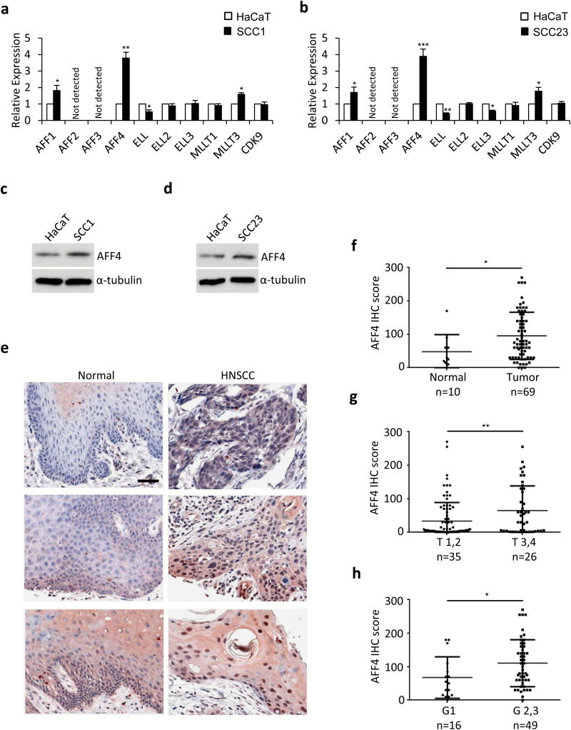
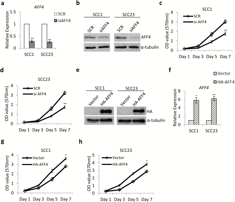
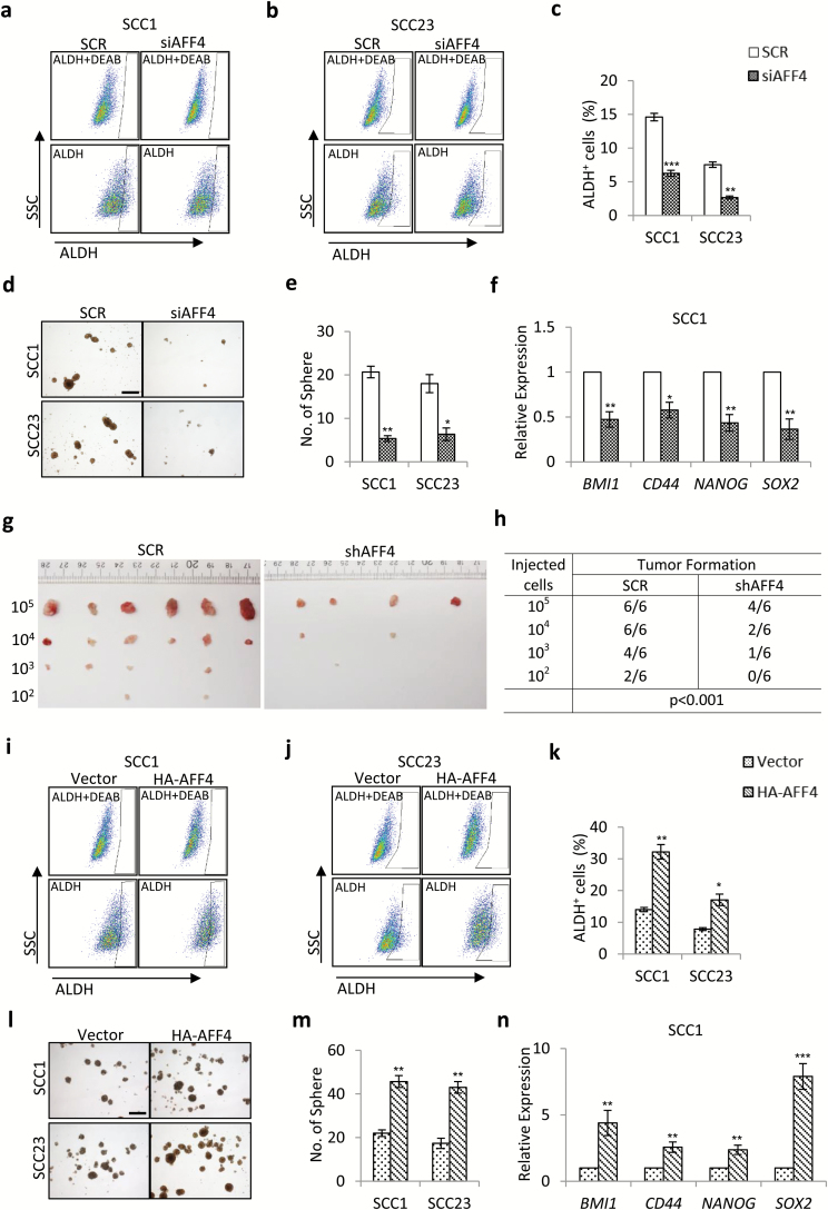
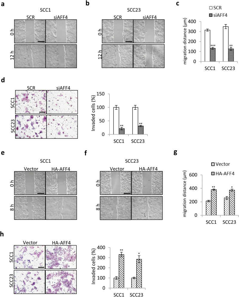
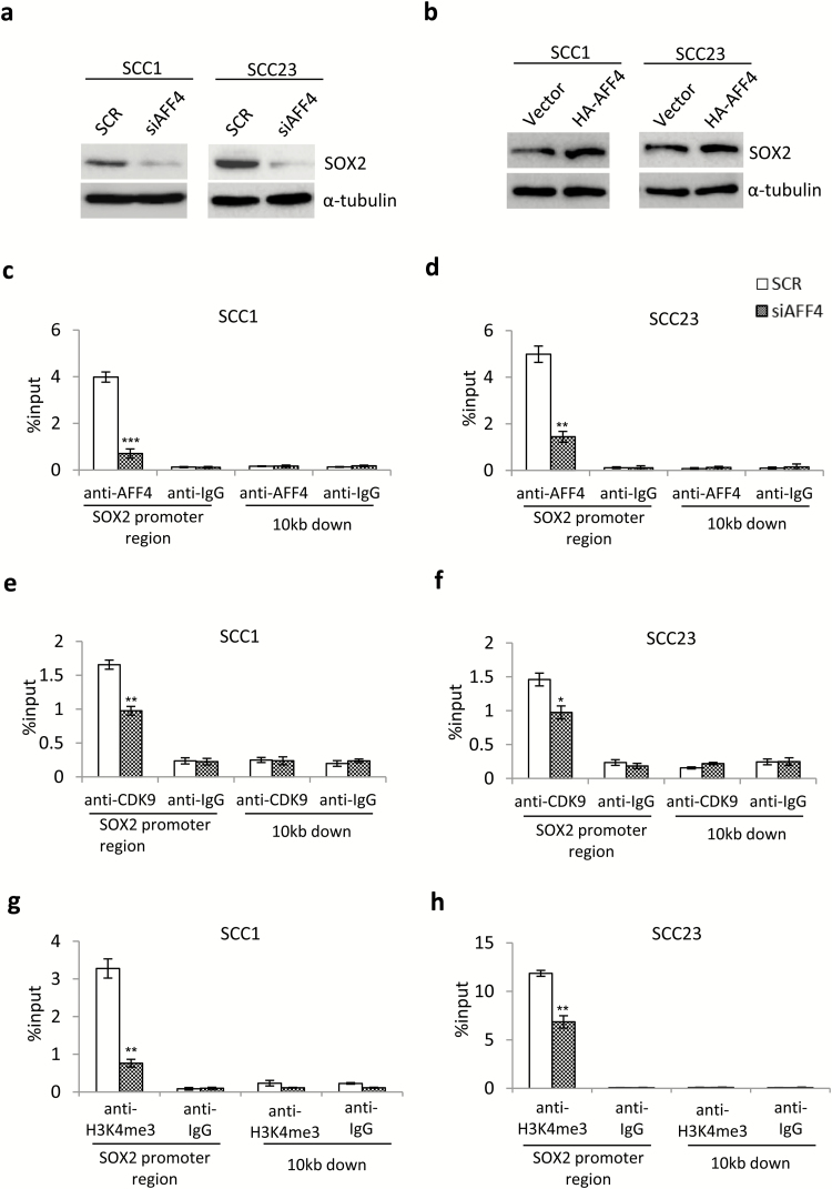
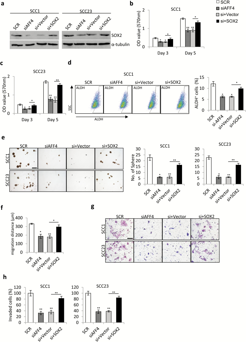
Super elongation complex (SEC) controls gene transcription by releasing Pol II from pausing. Previous studies have shown that dysfunction of SEC was associated with multiple human cancers, such as leukemia and breast cancer. However, the role of SEC in head and neck squamous cell carcinoma (HNSCC) development remains largely unknown. In this study, we found expression of AF4/FMR2 family member 4 (AFF4), the core component of SEC, was upregulated dramatically in HNSCC cell lines and tumor tissues. By using siRNA-mediated depletion and overexpression of AFF4, we demonstrated AFF4 promoted proliferation, migration and invasion of HNSCC cells. Moreover, we found AFF4 enhanced the aldehyde dehydrogenase (ALDH) activity and sphere formatting activity and was required for the tumor-initiation capacity of stem-like cells in HNSCC cell lines. Mechanistically, we found the role of AFF4 in regulation of HNSCC cell behaviors was mainly mediated by sex-determining region Y box2 (SOX2), a critical regulator involved in development of several human cancers. SOX2 expression changed in parallel with AFF4 expression in response to depletion and overexpression of AFF4, respectively. More importantly, overexpression of SOX2 rescued the inhibited proliferation, migration, invasion and ALDH activity induced by knockdown of AFF4 in HNSCC cells, at least in part. Collectively, our findings indicate AFF4 may serve as a biomarker and a potential target of therapies for patients with HNSCC.
Figures
Similar articles
-
C-Met pathway promotes self-renewal and tumorigenecity of head and neck squamous cell carcinoma stem-like cell.Oral Oncol. 2014 Jul;50(7):633-9. doi: 10.1016/j.oraloncology.2014.04.004. Epub 2014 May 15. Oral Oncol. 2014. PMID: 24835851 Review.
-
Regulation of Head and Neck Squamous Cancer Stem Cells by PI3K and SOX2.J Natl Cancer Inst. 2016 Sep 15;109(1):djw189. doi: 10.1093/jnci/djw189. Print 2017 Jan. J Natl Cancer Inst. 2016. PMID: 27634934 Free PMC article.
-
Super-Enhancer Driven LIF/LIFR-STAT3-SOX2 Regulatory Feedback Loop Promotes Cancer Stemness in Head and Neck Squamous Cell Carcinoma.Adv Sci (Weinh). 2024 Oct;11(40):e2404476. doi: 10.1002/advs.202404476. Epub 2024 Aug 29. Adv Sci (Weinh). 2024. PMID: 39206755 Free PMC article.
-
AFF4 facilitates melanoma cell progression by regulating c-Jun activity.Exp Cell Res. 2021 Feb 15;399(2):112445. doi: 10.1016/j.yexcr.2020.112445. Epub 2021 Jan 5. Exp Cell Res. 2021. PMID: 33417923
-
The Biological Significance of AFF4: Promoting Transcription Elongation, Osteogenic Differentiation and Tumor Progression.Comb Chem High Throughput Screen. 2024;27(10):1403-1412. doi: 10.2174/0113862073241079230920082056. Comb Chem High Throughput Screen. 2024. PMID: 37815186 Review.
Cited by
-
Butylidenephthalide Abrogates the Snail-Induced Cancer Stemness in Oral Carcinomas.Int J Mol Sci. 2022 May 31;23(11):6157. doi: 10.3390/ijms23116157. Int J Mol Sci. 2022. PMID: 35682836 Free PMC article.
-
FENDRR represses Bladder Cancer Cell Proliferation, Stemness, Migration, Invasion, and EMT Process by Targeting miR-18a-5p/AFF4 Axis.Biochem Genet. 2024 Nov 21. doi: 10.1007/s10528-024-10944-w. Online ahead of print. Biochem Genet. 2024. PMID: 39572480
-
The m6A Methylation-Regulated AFF4 Promotes Self-Renewal of Bladder Cancer Stem Cells.Stem Cells Int. 2020 Jul 2;2020:8849218. doi: 10.1155/2020/8849218. eCollection 2020. Stem Cells Int. 2020. PMID: 32676121 Free PMC article.
-
Targeting cancer stem cells in squamous cell carcinoma.Precis Clin Med. 2019 Sep;2(3):152-165. doi: 10.1093/pcmedi/pbz016. Epub 2019 Oct 1. Precis Clin Med. 2019. PMID: 31598386 Free PMC article. Review.
-
The Expression and Roles of the Super Elongation Complex in Mouse Cochlear Lgr5+ Progenitor Cells.Front Cell Neurosci. 2021 Oct 1;15:735723. doi: 10.3389/fncel.2021.735723. eCollection 2021. Front Cell Neurosci. 2021. PMID: 34658793 Free PMC article.
References
-
- Ferlay J., et al. (2010)Estimates of worldwide burden of cancer in 2008: GLOBOCAN 2008. Int. J. Cancer, 127, 2893–2917. - PubMed
-
- Global Burden of Disease Cancer Collaboration; Fitzmaurice C., et al. (2017)Global, regional, and national cancer incidence, mortality, years of life lost, years lived with disability, and disability-adjusted life-years for 32 cancer groups, 1990 to 2015: a systematic analysis for the global burden of disease study. JAMAOncol., 3, 524–548. - PMC - PubMed
-
- Carol R.B., et al. (1991)Human papillomavirus DNA sequences in cell lines derived from head and neck squamous cell carcinomas. Otolaryngol. Head Neck Surg., 104, 303–310. - PubMed
Publication types
MeSH terms
Substances
LinkOut - more resources
Full Text Sources
Other Literature Sources
Medical
