. 2018 Jun 5;90(11):6766-6772.
doi: 10.1021/acs.analchem.8b00655. Epub 2018 May 17.

Matrix-Independent Surface-Enhanced Raman Scattering Detection of Uranyl Using Electrospun Amidoximated Polyacrylonitrile Mats and Gold Nanostars

Matrix-Independent Surface-Enhanced Raman Scattering Detection of Uranyl Using Electrospun Amidoximated Polyacrylonitrile Mats and Gold Nanostars

Grace Lu et al. Anal Chem. .

Abstract

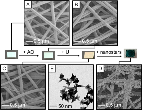
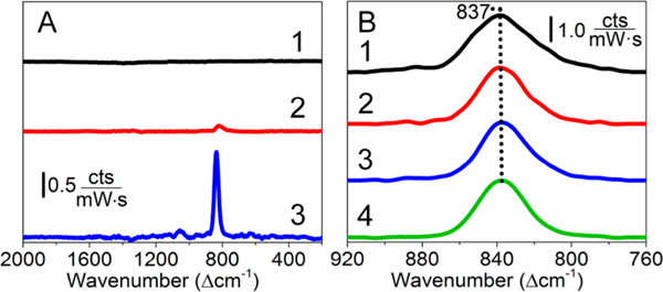
Reproducible detection of uranyl, an important biological and environmental contaminant, from complex matrixes by surface-enhanced Raman scattering (SERS) is successfully achieved using amidoximated-polyacrylonitrile (AO-PAN) mats and carboxylated gold (Au) nanostars. SERS detection of small molecules from a sample mixture is traditionally limited by nonspecific adsorption of nontarget species to the metal nanostructures and subsequent variations in both the vibrational frequencies and intensities. Herein, this challenge is overcome using AO-PAN mats to extract uranyl from matrixes ranging in complexity including HEPES buffer, Ca(NO3)2 and NaHCO3 solutions, and synthetic urine. Subsequently, Au nanostars functionalized with carboxyl-terminated alkanethiols are used to enhance the uranyl signal. The detected SERS signals scale with uranyl uptake as confirmed using liquid scintillation counting. SERS vibrational frequencies of uranyl on both hydrated and lyophilized polymer mats are largely independent of sample matrix, indicating less complexity in the uranyl species bound to the surface of the mats vs in solution. These results suggest that matrix effects, which commonly limit the use of SERS for complex sample analysis, are minimized for uranyl detection. The presented synergistic approach for isolating uranyl from complex sample matrixes and enhancing the signal using SERS is promising for real-world sample detection and eliminates the need of radioactive tracers and extensive sample pretreatment steps.


Figures

Figure 1.
Figure 1.
Overview of the isolation and detection of uranyl using AO-PAN mats and Au nanostars. Representative photographs and SEM images of the PAN mats (A) as fabricated (d = 101 ± 28 nm), after (B) AO functionalization (d =113 ± 22 nm), (C) uranyl uptake (d = 116 ± 24 nm), and (D) Au nanostar deposition are shown. In addition, (E) TEM images of the 6-MHA functionalized Au nanostars are shown (size = 59.8 ± 14.0 nm).
Figure 2.
Figure 2.
Confirmation of U uptake. (A) Deprotonation of AO groups as a function of pH. (B) Evaluation of uranyl sorption as a function of incubation time using AO-PAN mats and LSC. A standard kinetic model is used to fit the data (y = 8.5x/(4.43 + x)). Error bars = standard deviations of 2+ measurements. (C) Adsorbed U determined using LSC as a function of initial U concentration in 10 mM HEPES buffer as well as in HEPES buffer with 500 mg/L of Ca2+ or HCO3 or synthetic urine.
Figure 3.
Figure 3.
Evaluation of uranyl detectability using Raman microscopy. (A) Normal Raman spectra of 10 mM uranyl uptake from pH (1) 4 and (2) 6.8 solutions onto lyophilized AO-PAN mats. (B) Normal Raman spectra of (1) 1 and (2) 10 mM uranyl collected using hydrated and (3) 1 and (4) 10 mM uranyl using lyophilized AO-PAN mats. Spectra are collected using the following parameters: λex = 785 nm; tint= 50 s, and P = 55 mW, 5 averages; 50× objective; 10 mM HEPES was used (18 h incubation).
Figure 4.
Figure 4.
(A) Normal Raman spectra of 1 mM uranyl after uptake on hydrated (1) PAN and (2) AO-PAN mats as well as (3) a representative SERS spectrum of 10 μM uranyl after uptake on hydrated AO-PAN mats. (B) SERS spectra of 10 μM uranyl incubated with Au nanostars then deposited onto a (1) glass slide (ν¯=836.2±1.5cm1andΓ=35.2±1.5cm1), (2) PAN mat (ν¯=836.1±0.7cm1andΓ=30.9±1.0cm1), (3) AO-PAN mat (ν¯=837.1±0.5cm1andΓ=30.3±0.6cm1) and 10 μM uranyl (4) uptake on hydrated AO-PAN mats followed by addition of Au nanostars (ν¯=838.1±0.5cm1andΓ=30.3±1.0cm1). Collection conditions for normal Raman spectra are the same as in Figure 3. SERS collection parameters: P = 25 mW, tint = 30 s, 10× objective (hydrated) or P = 1.5 mW, tint = 50 s, 50× objective (lyophilized).
Figure 5.
Figure 5.
SERS spectra of 10 μM uranyl (pH 6.8) in (1) 10 mM HEPES, (2) 3.4 mM Ca2+, (3) 5 mM HCO32−, and (4) synthetic urine using (a) lyophilized and (B) hydrated AO-PAN mats and 6-MHA functionalized Au nanostars. SERS on (C) lyophilized and (D) hydrated mats vs mass U sorbed determined by LSC. Error bars represent noise in each individual measurement. The CH2 bending mode from 6-MHA is observed in lyophilized spectra and is centered at 817 cm−1. All other spectral features are from uranyl (Table 1). Collection same as Figure 4.
Scheme 1.
Scheme 1.
Proposed Pathway of Uranyl Uptake on AO-PAN Mats from Solution via Hydroxylamine Coordination to Uranyl with (A) All Aqua and (B) Two Aqua and One Hydroxyl Ligands and (C) Coordination with Carboxylic Acid from 6-MHA Functionalized Au Nanostars

References

    1. Blake JM; Avasarala S; Artyushkova K; Ali A-MS; Brearley AJ; Shuey C; Robinson WP; Nez C; Bill S; Lewis J; Hirani C; Pacheco JSL; Cerrato JM Environ. Sci. Technol 2015, 49, 8506–8514. - PubMed
    1. U.S. Environmental Protection Agency. Pacific Southwest, Region; 9, 2015.
    1. Bernhard G; Geipel G; Brendler V; Nitsche HJ Alloys Compd. 1998, 271–273, 201–205.
    1. Berto S; Crea F; Daniele PG; Gianguzza A; Pettignano A; Sammartano S Coord. Chem. Rev. 2012, 256, 63–81.
    1. Rout S; Ravi PM; Kumar A; Tripathi RM J. Radioanal. Nucl. Chem. 2015, 303, 2193–2203.

Publication types

LinkOut - more resources