. 2018 Jul 1;29(13):1664-1674.
doi: 10.1091/mbc.E18-02-0129. Epub 2018 May 9.

The kinase domain of CK1 enzymes contains the localization cue essential for compartmentalized signaling at the spindle pole

Affiliations

The kinase domain of CK1 enzymes contains the localization cue essential for compartmentalized signaling at the spindle pole

Zachary C Elmore et al. Mol Biol Cell. .

Abstract

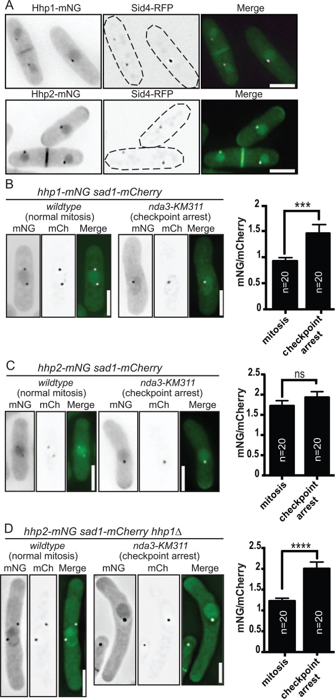
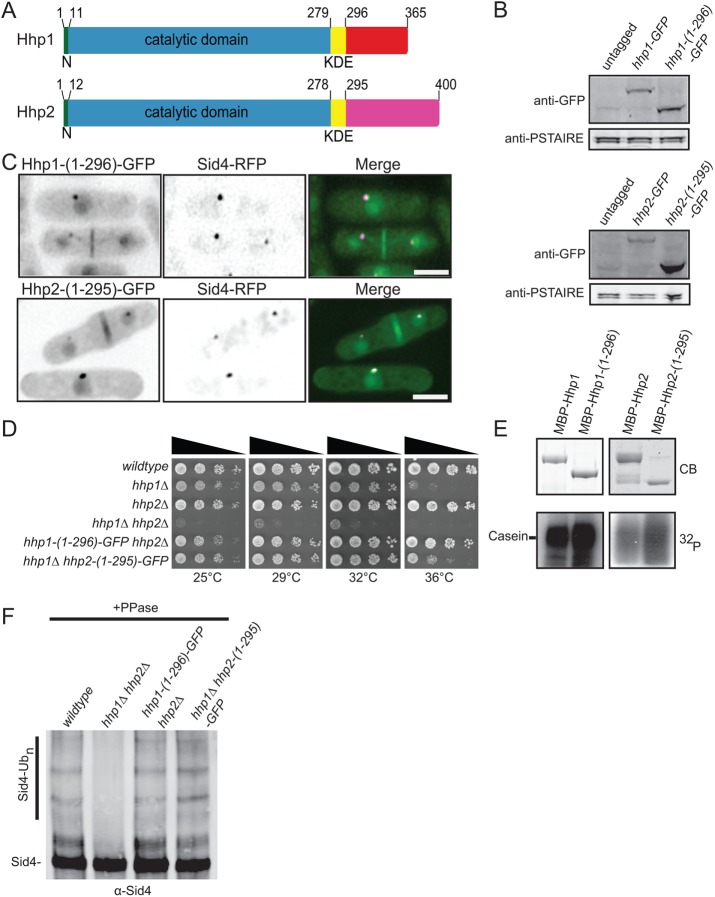
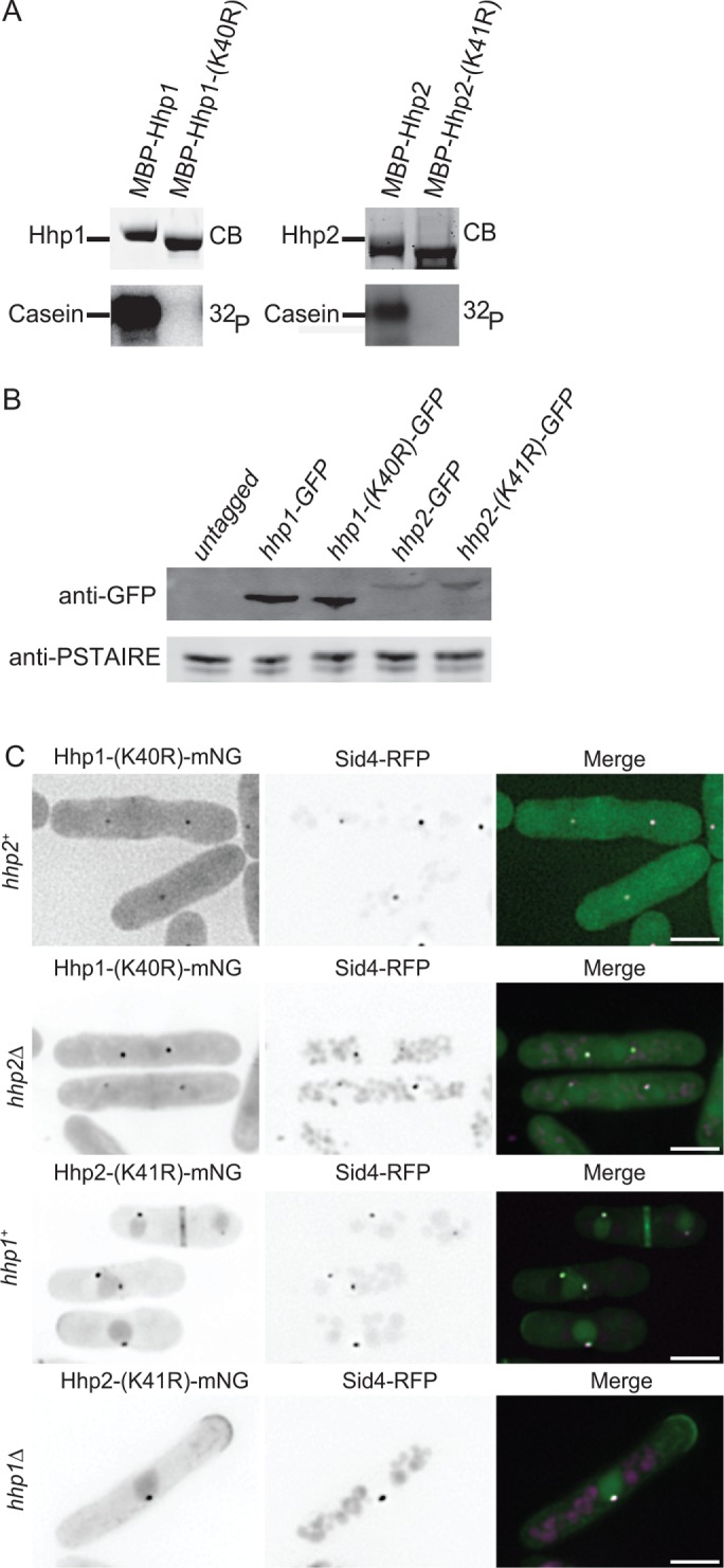
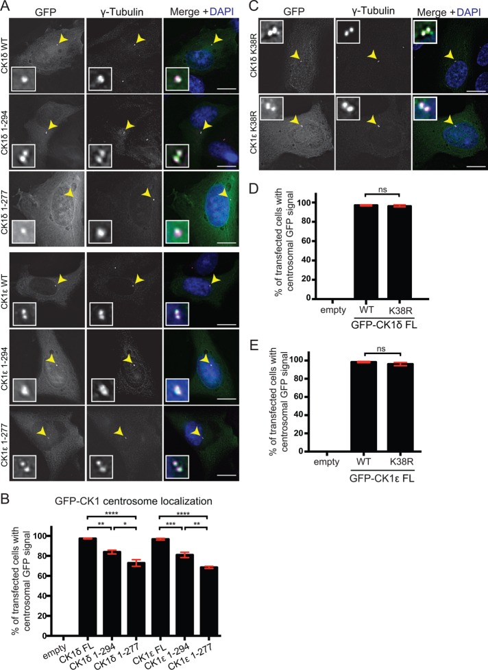
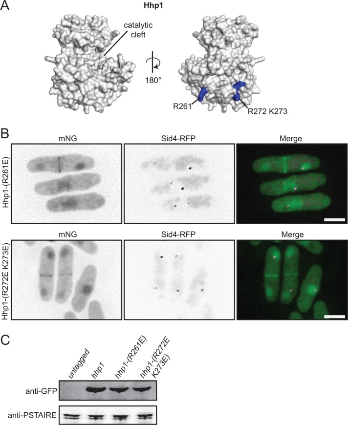
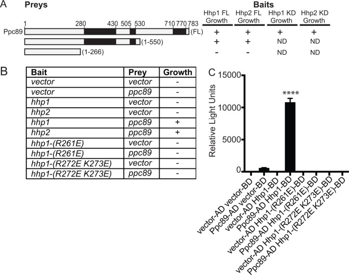
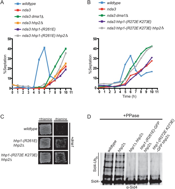
CK1 protein kinases contribute to multiple biological processes, but how they are tailored to function in compartmentalized signaling events is largely unknown. Hhp1 and Hhp2 (Hhp1/2) are the soluble CK1 family members in Schizosaccharomyces pombe. One of their functions is to inhibit the septation initiation network (SIN) during a mitotic checkpoint arrest. The SIN is assembled by Sid4 at spindle pole bodies (SPBs), and though Hhp1/2 colocalize there, it is not known how they are targeted there or whether their SPB localization is required for SIN inhibition. Here, we establish that Hhp1/2 localize throughout the cell cycle to SPBs, as well as to the nucleus, cell tips, and division site. We find that their catalytic domains but not their enzymatic function are used for SPB targeting and that this targeting strategy is conserved in human CK1δ/ε localization to centrosomes. Further, we pinpoint amino acids in the Hhp1 catalytic domain required for SPB interaction; mutation of these residues disrupts Hhp1 association with the core SPB protein Ppc89, and the inhibition of cytokinesis in the setting of spindle stress. Taken together, these data have enabled us to define a molecular mechanism used by CK1 enzymes to target a specific cellular locale for compartmentalized signaling.


Figures

FIGURE 1:
FIGURE 1:
Intracellular localization patterns of Hhp1/2. (A) Live-cell imaging of Hhp1-mNG and Hhp2-mNG with Sid4-RFP. (B–D) Hhp1-mNG (B) and Hhp2-mNG (C, D) were imaged in prometaphase-arrested and wild-type mitotic sad1-mCherry or sad1-mCherry hhp1∆ cells. Representative inverted grayscale images are shown in the left panels with quantitation to the right. mCh, mCherry. Scale bars: 5 µm. Values are represented as mNG/mCherry intensity ratios. ***, p < 0.005, ****, p < 0.001 determined using Student’s t test. ns, not significant. Error bars represent SEM.
FIGURE 2:
FIGURE 2:
The C-termini of Hhp1/2 are dispensable for their functions. (A) Schematic diagrams of Hhp1/2 with relative positions of N-terminal extensions (N, green), kinase domains (blue), kinase domain extensions (KDE, yellow), and unrelated C-termini in red or purple indicated, drawn to scale. (B) Anti-GFP immunoblots of whole-cell extracts prepared from untagged and the indicated GFP-tagged strains. Anti-PSTAIRE (Cdc2) immunoblots served as protein loading controls. (C) Live-cell imaging of endogenously tagged Hhp1-(1-296)-GFP and Hhp2-(1-295)-GFP with Sid4-RFP. Scale bars: 5 µm. (D) Serial 10-fold dilutions of the indicated strains were spotted on YE plates and incubated at the indicated temperatures. (E) In vitro kinase assays of recombinant MBP-Hhp1, MBP-Hhp1-(1-296), MBP-Hhp2, and MBP-Hhp2-(1-295) detected by Coomassie blue (CB) staining of SDS–PAGE gels, with casein as substrate. Phosphorylated casein was detected by autoradiography (32P). (F) Sid4 from the indicated strains was immunoprecipitated from denatured cell lysates, treated with phosphatase, and visualized by immunoblotting.
FIGURE 3:
FIGURE 3:
Kinase activity of Hhp1/2 is not required for SPB localization. (A) In vitro kinase assays of recombinant MBP-Hhp1-(K40R) and MBP-Hhp2-(K41R) detected by CB staining of SDS–PAGE gels, with casein as substrate. Phosphorylated casein was detected by autoradiography. (B) Anti-GFP immunoblot of whole-cell extracts prepared from untagged and the indicated GFP-tagged strains. Anti-PSTAIRE antibody served as loading control for lysates. (C) Live-cell imaging of endogenously tagged Hhp1-(K40R)-mNG with Sid4-RFP in wildtype and hhp2∆ cells along with Hhp2-(K41R)-mNG with Sid4-RFP in wildtype and hhp1∆ cells. Scale bars: 5 µm.
FIGURE 4:
FIGURE 4:
The centrosomal targeting information of CK1δ/ε is located within the kinase domain. RPE-1 cells were transiently transfected with GFP N-terminal fusions to full length or truncations of CK1δ and CK1ε. Cells were fixed with 100% methanol and stained with γ-tubulin (magenta) and DAPI (blue). (A) Localization of full-length protein and truncation mutants in RPE-1 cells. (B) Quantification of the colocalization between γ-tubulin and full-length proteins or truncation mutants. (C) Localization of full-length GFP-CK1δ and GFP-CK1ε catalytically inactive mutants. (D, E) Quantification of the colocalization between γ-tubulin and GFP-CK1δ (D) or GFP-CK1ε (E) wild type or catalytically inactive mutants. For B, D, and E, 100 cells per experiment. n = 3. *, p < 0.05, **, p < 0.01, ***, p < 0.005, ****, p < 0.001, p values determined using ANOVA; ns, not significant. Error bars represent SEM. Scale bars: 15 μm.
FIGURE 5:
FIGURE 5:
Residues at the base of the Hhp1 catalytic domain are critical for SPB localization. (A) Homology model of Hhp1 catalytic domain generated from Phyre2 software and visualized with MacPymol. Residues critical for Hhp1 SPB localization are in blue. (B) Live-cell imaging of endogenously tagged Hhp1-(R261E)-mNG and Hhp1-(R272E K273E)-mNG with Sid4-RFP. Scale bars: 5 μm. (C) Anti-GFP immunoblot of whole-cell extracts prepared from the indicated strains. Anti-PSTAIRE immunoblots served as protein loading controls.
FIGURE 6:
FIGURE 6:
Basic residues within the kinase domain of Hhp1 are critical for interaction with the SPB protein Ppc89. (A, B) S. cerevisiae strain PJ69-4A was cotransformed with plasmids expressing hhp1/2 or their variants, hhp1-(1-296), or hhp2-(1-295) and indicated sections of ppc89. KD = kinase domain. Black boxes in A indicate regions of predicted coiled-coil. Transformants were scored for growth on −Trp −Leu −His plates supplemented with 5 mM 3-AT. Pluses indicate strong growth and minuses indicate no growth. (C) Bar graph shows β-galactosidase activity of the indicated bait and prey plasmids tested for growth in B (represented in relative light units). Each assay was performed in triplicate. ****, p < 0.001 determined using ANOVA. Error bars represent SEM.
FIGURE 7:
FIGURE 7:
Hhp1 SPB localization is required for mitotic checkpoint function. (A, B) The indicated strains were synchronized in S phase with hydroxyurea and shifted to 19°C to activate the spindle checkpoint, and septation indices were measured periodically for 9 h. (C) Overexpression of dma1 from the nmt41 promoter in wild type, hhp1-(R261E) hhp2∆, or hhp1-(R272E K273E) hhp2∆ mutant cells. Growth of the transformants was observed on agar plates in the presence (repression) or absence (derepression) of thiamine. (D) Sid4 from the indicated strains was immunoprecipitated from denatured cell lysates, treated with phosphatase, and visualized by immunoblotting.

Similar articles

Cited by

References

    1. Agostinis P, Pinna LA, Meggio F, Marin O, Goris J, Vandenheede JR, Merlevede W. (1989). A synthetic peptide substrate specific for casein kinase I. FEBS Lett , 75–78. - PubMed
    1. Babu P, Bryan JD, Panek HR, Jordan SL, Forbrich BM, Kelley SC, Colvin RT, Robinson LC. (2002). Plasma membrane localization of the Yck2p yeast casein kinase 1 isoform requires the C-terminal extension and secretory pathway function. J Cell Sci , 4957–4968. - PubMed
    1. Babu P, Deschenes RJ, Robinson LC. (2004). Akr1p-dependent palmitoylation of Yck2p yeast casein kinase 1 is necessary and sufficient for plasma membrane targeting. J Biol Chem , 27138–27147. - PubMed
    1. Bahler J, Wu JQ, Longtine MS, Shah NG, McKenzie A, 3rd, Steever AB, Wach A, Philippsen P, Pringle JR. (1998). Heterologous modules for efficient and versatile PCR-based gene targeting in Schizosaccharomyces pombe. Yeast , 943–951. - PubMed
    1. Bhattacharyya RP, Remenyi A, Good MC, Bashor CJ, Falick AM, Lim WA. (2006). The Ste5 scaffold allosterically modulates signaling output of the yeast mating pathway. Science , 822–826. - PubMed

Publication types