. 2018 Jul 1;29(13):1542-1554.
doi: 10.1091/mbc.E17-04-0248. Epub 2018 May 9.

The E3 ubiquitin ligase UBR5 regulates centriolar satellite stability and primary cilia

Affiliations

The E3 ubiquitin ligase UBR5 regulates centriolar satellite stability and primary cilia

Robert F Shearer et al. Mol Biol Cell. .

Erratum in

  • Correction.
    [No authors listed] [No authors listed] Mol Biol Cell. 2022 Feb 1;33(2):cor1. doi: 10.1091/mbc.E17-04-0248-corr. Mol Biol Cell. 2022. PMID: 35041470 Free PMC article. No abstract available.

Abstract

Primary cilia are crucial for signal transduction in a variety of pathways, including hedgehog and Wnt. Disruption of primary cilia formation (ciliogenesis) is linked to numerous developmental disorders (known as ciliopathies) and diseases, including cancer. The ubiquitin-proteasome system (UPS) component UBR5 was previously identified as a putative positive regulator of ciliogenesis in a functional genomics screen. UBR5 is an E3 ubiquitin ligase that is frequently deregulated in tumors, but its biological role in cancer is largely uncharacterized, partly due to a lack of understanding of interacting proteins and pathways. We validated the effect of UBR5 depletion on primary cilia formation using a robust model of ciliogenesis, and identified CSPP1, a centrosomal and ciliary protein required for cilia formation, as a UBR5-interacting protein. We show that UBR5 ubiquitylates CSPP1, and that UBR5 is required for cytoplasmic organization of CSPP1-comprising centriolar satellites in centrosomal periphery, suggesting that UBR5-mediated ubiquitylation of CSPP1 or associated centriolar satellite constituents is one underlying requirement for cilia expression. Hence, we have established a key role for UBR5 in ciliogenesis that may have important implications in understanding cancer pathophysiology.


Figures

FIGURE 1:
FIGURE 1:
Centriolar satellites as regulators of centrosomal function. (A) Centriolar satellites localize to the pericentriolar space surrounding the centrioles. Cilium and centrioles stained with an antibody to glutamylated-tubulin (red), and satellites stained with antibody to PCM1 (green). Nuclear marker Hoechst33258 (blue). High-powered inset fields are indicated by a dashed box. Confocal imaging, bar = 10 µm. (B) Centriolar satellites are required for shuttling of centrosomal proteins to the basal body from which the ciliary axoneme extends during G1 phase (Lopes et al., 2011), and act in the assembly of proteins during centriole duplication in S phase (Hori and Toda, 2017).
FIGURE 2:
FIGURE 2:
UBR5 depletion disrupts centriolar satellite stability and primary cilia formation in hTERT-RPE1 cells. (A) Schematic describing ciliogenesis assay. (B) Depletion of UBR5 attenuates primary cilia formation in and causes dispersion of centriolar satellites in RPE1 cells. Cilium and basal body stained with antibody to glutamylated tubulin (red), satellites stained with antibody to PCM1 (green), and nucleus marked with Hoechst33258 (blue). A similar phenotype is observed for depletion of PCM1. High-powered inset fields are indicated by a dashed box. Transfections performed 72 h before imaging. Data representative of two independent experiments, with 150 cells counted per condition per experiment. Confocal imaging, bar = 10 µm. High magnification of region of interest (ROI) bar = 2 µm. (C) Quantitation of data depicted in B shows the strong penetrance of siUBR5 and siPCM1 phenotype. Blue and red bars show percentage of cells with distinct or dispersed satellites, respectively. Error bars = SEM. (D) Immunoblot showing siRNA efficacy in RPE1 cells used for staining in C. (E) Coimmunoprecipitation of UBR5 and Western blotting for potential interactions with PCM1, MIB1, and γ-tubulin (a marker of the centrosome). Transfections performed 48 h before imaging. Data representative of two experiments.
FIGURE 3:
FIGURE 3:
Coexpression of UBR5 gene. (A) Waterfall plot showing global correlation of gene expression against UBR5 expression in NCI-60 cancer cell line panel, obtained via CellMiner (Reinhold et al., 2012). Cutoff set at Pearson correlation of 0.254. Putative ciliary proteins are indicated, with putative top tier cilia localizing proteins labeled and indicated with larger red dots; second tier cilia localizing proteins are also labeled and shown with larger gray dots. Putative cilia localizing proteins based on cilia proximity labeling data (Mick et al., 2015). (B) Structure of CSPP1 protein isoforms. Note that the extra 294 amino acid N-terminal region of CSPP-L is the binding region of the selected CSPP1 antibody. N-Degron motif shown in red. Coiled-coil domains are shown with a black box. Proline-rich domains are shown with a white box. Glutamate-rich domains are shown with a white striped box.
FIGURE 4:
FIGURE 4:
UBR5 binds and polyubiquitylates CSPP-L. (A) Schematic showing the structure of UBR5 protein and the position of known functional domains and amino acid changes of mutants. (B) Coimmunoprecipitation and immunoblot analysis of GFP-UBR5 (and functional mutants) transfected into HEK293T cells. GFP-UBR5 coimmunoprecipitates endogenous ubiquitylated CSPP-L. Covalent modification of CSPP-L is quantified by an integrated density plot showing relative size shift. Note the reduction of modified CSPP-L binding HECT* mutant. GFP-UBR5 and mutants are predicted to be approximately 338 kDa. Transfections performed 24 h before harvest. WCE, whole cell extract; NB, nonbound fraction (4% of input); IP, immunoprecipitation. Data are representative of two experiments. (C) Coimmunoprecipitation and immunoblot analysis of GFP-CSPP-L transfected into HEK293T cells. GFP-CSPP-L binds endogenous UBR5. Endogenous Ub is detected covalently bound to GFP-CSPP-L. The far right panel shows coimmunoprecipitation performed under denaturing conditions. GFP-CSPP-L is predicted to be approximately 170 kDa. Data are representative of two experiments.
FIGURE 5:
FIGURE 5:
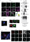
UBR5 interacts with CSPP-L at the centrosome. (A) Left to right, CSPP-L interacts with UBR5 in large foci adjacent to the nucleus (arrows). CSPP-L interacts with Ub in large foci adjacent to the nucleus (arrows). Interaction between UBR5 and Ub in HEK293T cells shows nuclear and cytoplasmic localization, with large foci of UBR5 and Ub in the nucleus. Untransfected cells show no visible signal. Nuclear marker is H2B-mCherry (magenta). High-powered inset fields are indicated by a dashed box (bar = 30 µm) and schematic of BiFC analysis for protein–protein interactions included. Data are representative of two experiments. (B) Immunoblot data showing correct production of fusion proteins in BiFC assay. Expected fusion protein sizes are V2-CSPP-L 152 kDa, V1-UBR5 329 kDa, V1-Ub 28 kDa, and V2-Ub 19 kDa. (C) CSPP-L/Ub interacting pairs colocalize with PCM1 (centriolar satellite marker) in hTERT-RPE1 cells. Bar = 20 µm. High magnification of ROI bar = 2 µm. (D) High-level expression of CSPP-L/Ub BiFC vectors shows CSPP-L and Ub interaction is confined to the centriolar satellites, despite strong detection of CSPP-L at the MTs in HEK293T cells. Region of interest (ROI) 1 shows relatively high BiFC pair expression, ROI 2 shows relatively low BiFC pair expression, and ROI 3 shows no BiFC pair expression. Bar = 10 µm. High magnification of ROI bar = 2 µm.
FIGURE 6:
FIGURE 6:
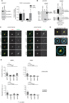
UBR5 maintains CSPP-L at the centrosome/centriolar satellites, and is required for centriolar satellite stability. (A) Depletion of UBR5 by shRNA in HEK293T cells does not decrease CSPP-L levels. shRNA induced with 1 µg/ml doxycycline 48 h before harvest. Data summary represents results from four independent experiments; n = 6 replicates per experiment. (B) Immunoprecipitation of GFP-CSPP-L coimmunoprecipitates CSPP-L–bound lysine-48 (K48) linked Ub chains and lysine-63 (K63) linked Ub chains. Transfections performed 24 h before harvest. (C) Depletion of UBR5 (but not PCM1) causes dispersion of centrosomal CSPP-L in RPE1 cells. (D) Centrosome/centriolar satellite mask. (E) Quantitation of loss of CSPP-L/PCM1 at the centrosome according to the mask described in D. At least 30 cells were scored per sample and statistical analysis performed using two-tailed t test.

Similar articles

Cited by

References

    1. Akizu N, Silhavy JL, Rosti RO, Scott E, Fenstermaker AG, Schroth J, Zaki MS, Sanchez H, Gupta N, Kabra M, et al (2014). Mutations in CSPP1 lead to classical Joubert syndrome. Am J Hum Genet , 80–86. - PMC - PubMed
    1. Asiedu M, Wu D, Matsumura F, Wei Q. (2009). Centrosome/spindle pole-associated protein regulates cytokinesis via promoting the recruitment of MyoGEF to the central spindle. Mol Biol Cell , 1428–1440. - PMC - PubMed
    1. Bangs FK, Schrode N, Hadjantonakis AK, Anderson KV. (2015). Lineage specificity of primary cilia in the mouse embryo. Nat Cell Biol , 113–122. - PMC - PubMed
    1. Barretina J, Caponigro G, Stransky N, Venkatesan K, Margolin AA, Kim S, Wilson CJ, Lehar J, Kryukov GV, Sonkin D, et al (2012). The Cancer Cell Line Encyclopedia enables predictive modelling of anticancer drug sensitivity. Nature , 603–607. - PMC - PubMed
    1. Bjellqvist B, Hughes GJ, Pasquali C, Paquet N, Ravier F, Sanchez JC, Frutiger S, Hochstrasser D. (1993). The focusing positions of polypeptides in immobilized pH gradients can be predicted from their amino acid sequences. Electrophoresis , 1023–1031. - PubMed

Publication types

LinkOut - more resources