Review
. 2018;113(7):e180040.
doi: 10.1590/0074-02760180040. Epub 2018 May 7.

Peeling the onion: the outer layers of Cryptococcus neoformans

Affiliations
Review

Peeling the onion: the outer layers of Cryptococcus neoformans

Daniel P Agustinho et al. Mem Inst Oswaldo Cruz. 2018.

Abstract

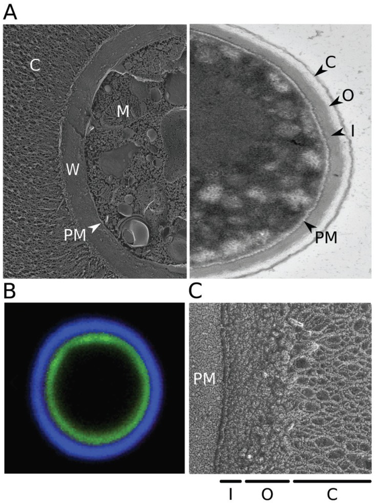
Cryptococcus neoformans is an opportunistic fungal pathogen that is ubiquitous in the environment. It causes a deadly meningitis that is responsible for over 180,000 deaths worldwide each year, including 15% of all AIDS-related deaths. The high mortality rates for this infection, even with treatment, suggest a need for improved therapy. Unique characteristics of C. neoformans may suggest directions for drug discovery. These include features of three structures that surround the cell: the plasma membrane, the cell wall around it, and the outermost polysaccharide capsule. We review current knowledge of the fundamental biology of these fascinating structures and highlight open questions in the field, with the goal of stimulating further investigation that will advance basic knowledge and human health.


Figures

None
(A) Electron micrographs of Cryptococcus neoformans. Left, quick-freeze deep-etch image of cells grown in capsule-inducing conditions (as in Haynes et al. 2011); right, transmission image of cells grown in rich medium (YPD), which yields only thin capsules. C: capsule; W: cell wall; PM: plasma membrane; M: mitochondrion; O: outer cell wall layer; I: inner cell wall layer. (B) Fluorescent micrograph highlighting the cell wall and capsule. Cryptococcal cells were induced to form capsule and stained with florescein (green) to label the cell wall and monoclonal antibody 2H1 (blue, generously provided by Arturo Casadevall) to label the capsule, as in Pierini and Doering (2001). (C) Quick-freeze deep-etch image highlighting the two layers of the cell wall.

References

    1. Agustinho DP, Nosanchuk JD. Functions of fungal melanins. In: Reference module in life sciences. 2017. Available from: http://www.sciencedirect.com/science/article/pii/B9780128096338120916.
    1. Alspaugh JA. Virulence mechanisms and Cryptococcus neoformans pathogenesis. Fungal Genet Biol. 2015;78:55–58. - PMC - PubMed
    1. Araújo GRS, Freitas GJC, Fonseca FL, Leite PEC, Rocha GM, de Souza W, et al. The environmental yeast Cryptococcus liquefaciens produces capsular and secreted polysaccharides with similar pathogenic properties to those of C. neoformans . Sci Rep. 2017;4:67–68. - PMC - PubMed
    1. Araújo GS, Fonseca FL, Pontes B, Torres A, Cordero RJB, Zancop-Oliveira RM, et al. Capsules from pathogenic and non-pathogenic Cryptococcus spp. manifest significant differences in structure and ability to protect against phagocytic cells. PLoS ONE. 2012;7(1):e29561. - PMC - PubMed
    1. Bahn YS, Jung KW. Stress signaling pathways for the pathogenicity of Cryptococcus . Eukaryot Cell. 2013;12(12):1564–1577. - PMC - PubMed

MeSH terms