. 2018 Dec;68(6):2118-2129.
doi: 10.1002/hep.30087. Epub 2018 Nov 1.

Binding of Free and Immune Complex-Associated Hepatitis C Virus to Erythrocytes Is Mediated by the Complement System

Affiliations

Binding of Free and Immune Complex-Associated Hepatitis C Virus to Erythrocytes Is Mediated by the Complement System

Kazi Abdus Salam et al. Hepatology. 2018 Dec.

Abstract

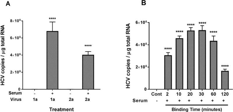
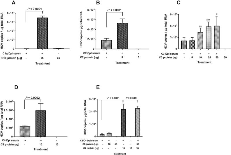
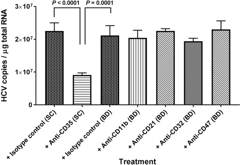
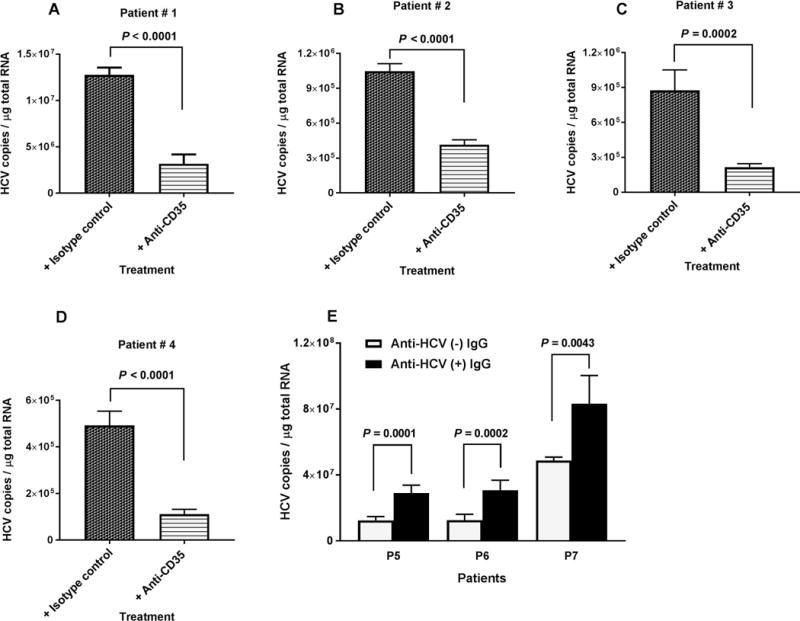
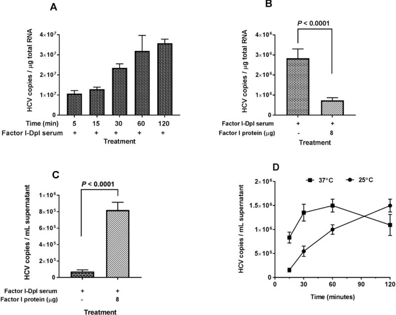
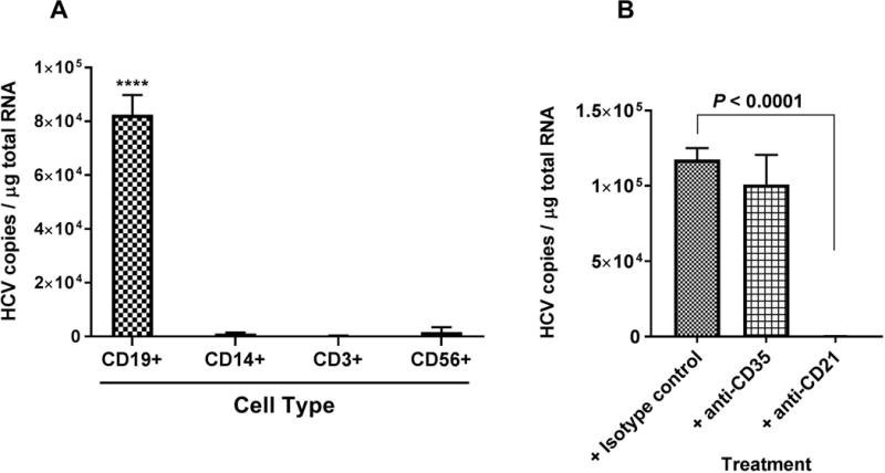
Erythrocytes bind circulating immune complexes (ICs) and facilitate IC clearance from the circulation. Chronic hepatitis C virus (HCV) infection is associated with IC-related disorders. In this study, we investigated the kinetics and mechanism of HCV and HCV-IC binding to and dissociation from erythrocytes. Cell culture-produced HCV was mixed with erythrocytes from healthy blood donors, and erythrocyte-associated virus particles were quantified. Purified complement proteins, complement-depleted serum, and complement receptor antibodies were used to investigate complement-mediated HCV-erythrocyte binding. Purified HCV-specific immunoglobulin G (IgG) from a chronic HCV-infected patient was used to study complement-mediated HCV-IC/erythrocyte binding. Binding of HCV to erythrocytes increased 200- to 1,000-fold after adding complement active human serum in the absence of antibody. Opsonization of free HCV occurred within 10 minutes, and peak binding to erythrocytes was observed at 20-30 minutes. Complement protein C1 was required for binding, whereas C2, C3, and C4 significantly enhanced binding. Complement receptor 1 (CR1, CD35) antibodies blocked the binding of HCV to erythrocytes isolated from chronically infected HCV patients and healthy blood donors. HCV-ICs significantly enhanced complement-mediated binding to erythrocytes compared to unbound HCV. Dissociation of complement-opsonized HCV from erythrocytes depended on the presence of Factor I. HCV released by Factor I bound preferentially to CD19+ B cells compared to other leukocytes. Conclusion: These results demonstrate that complement mediates the binding of free and IC-associated HCV to CR1 on erythrocytes and provide a mechanistic rationale for investigating the differential phenotypic expression of HCV-IC-related disease.


Conflict of interest statement

Potential conflict of interest: Nothing to report.

Figures

FIG. 1
FIG. 1
(A) Factors in serum are required for HCV genotype 1a and 2a binding to erythrocytes. Three milliliters medium containing HCV virus as indicated [HCV1a (H77s), 3 × 10⁷ copies total; HCV2a (JFH1), 2 × 10⁸ copies total] were incubated in the absence (100 μL medium only) or in the presence of serum (100 μL) at 25°C for 30 minutes. Then 2 mL of erythrocytes (2.5 × 10⁸ cells/mL) in complete RPMI medium were added to the reaction mixture and incubated further at 25°C for 15 minutes. Finally, a final concentration of 20 mM EDTA was added to stop the reaction, and the reaction mixture was immediately cooled for 5 minutes in an ice bath. Erythrocytes were collected by centrifugation and washed three times with 1× PBS, pH7.4. Total RNA isolation from erythrocytes and quantification of HCV RNA were performed as described in the Methods section. HCV associated with erythrocytes was significantly higher for both genotypes in the presence of serum. (B) Kinetic profiles for HCV binding to erythrocytes. For the optimal binding time, 3 mL medium containing HCV1a virus (1.5 × 10⁷ copies total) were treated in the presence of serum (100 μL) at 25°C for 30 minutes. The erythrocytes (5 × 10⁸ cells total) in 2 mL RPMI medium were then added and the incubation continued at 25°C with variable times as stated. A control (Cont) experiment without serum (100 μL medium only) was performed for each assay, and incubated at 25°C for the longest time point as indicated. After adding EDTA to a final concentration of 20 mM to stop the reaction, and the reaction mixture was immediately cooled for 5 minutes in an ice bath. Erythrocytes were collected by centrifugation and washed three times with 1× PBS, pH7.4. Total RNA isolation from erythrocytes and quantification of HCV RNA were performed as described in the Methods section. HCV associated with erythrocytes was significantly higher at each time point in the presence of serum. Each value represents the mean ± standard deviation of six determinations. The data are representative of two independent experiments using erythrocytes from at least two different healthy donors. **** P < 0.0001, compared to absence of serum; control (serum absent).
FIG. 2
FIG. 2
Purified complement proteins C1q, C2, C3, and C4 added to the indicated complement-depleted serum can restore HCV binding to erythrocytes. (A-E) Three milliliters of medium containing HCV1a virus (1 to 3 × 10⁷ genomic copies total) were treated with 100 μL of the indicated complement-depleted serum sample, complement-depleted serum sample plus the indicated purified complement protein, or purified complement protein only. After 30 minutes incubation at 25°C, 2 mL of erythrocytes (5 × 10⁸ cells total) in complete RPMI medium were added to the reaction mixture and incubated further at 25°C for 15 minutes. After adding EDTA to a final concentration of 20 mM, the erythrocytes were collected by centrifugation and washed three times with 1× PBS, pH7.4. Total RNA isolation from erythrocytes and quantification of HCV RNA were performed as described in Methods section. Each value represents the mean ± standard deviation of six determinations. The data are representative of two independent experiments using erythrocytes from at least two different healthy donors. Dpl, depleted.
FIG. 3
FIG. 3
Antibodies against CR1 (anti-CD35) block binding of HCV to erythrocytes. The erythrocytes (5 × 10⁸ cells total) in 2 mL of RPMI medium were mixed with 6 μg antibodies as stated and incubated at 25°C for 30 minutes. A mixture of complement opsonized HCV1a virus particles (pre-incubation at 25°C for 30 minutes, 1.5 × 10⁷ copies total) were added and incubated further at 25°C for 15 minutes. After adding EDTA to a final concentration of 20 mM, the erythrocytes were collected by centrifugation and washed three times with 1× PBS, pH7.4. Total RNA isolation from erythrocytes and quantification of HCV RNA were performed as described in the Methods section. Each value represents the mean ± standard deviation of six determinations. The data are representative of two independent experiments using erythrocytes from at least two different healthy donors. SC, Santa Cruz; BD, BD Biosciences.
FIG. 4
FIG. 4
Effects of anti-CD35 antibody and HCV specific polyclonal antibodies on HCV binding to erythrocytes. (A-D) Erythrocytes from four chronically infected HCV patients in 2 mL of RPMI medium (5 × 10⁸ cells total) were incubated with 4 to 6 μg antibodies as indicated at 25°C for 30 minutes, followed by adding 3 mL of complement opsonized HCV1a virus (pre-incubation for 30 minutes at 25°C, 1 to 1.5 × 10⁷ genomic copies total) and incubating further at 25°C for 15 minutes. (E) Three milliliters of HCV1a (1 × 10⁷ genomic copies total) were incubated with heat aggregated IgG (100 μg/mL) as indicated at 25°C for 30 minutes, followed by mixing with 100 μL serum and incubating at 25°C for 30 minutes. Then, 2 mL erythrocytes (2.5 × 10⁸ cells per mL), isolated from three chronically infected HCV patients were added. The reaction was carried out at room temperature (25°C) for 15 minutes. After adding EDTA to a final concentration of 20 mM, the erythrocytes were collected by centrifugation and washed three times with 1× PBS, pH7.4. Total RNA isolation from erythrocytes and quantification of HCV RNA were performed as described in the Methods section. Each value represents the mean ± standard deviation of six determinations.
FIG. 5
FIG. 5
Effects of Factor I on HCV association with erythrocytes. (A) Three milliliters of HCV1a virus (1 × 10⁷ genomic copies total) were incubated with 50 μL Factor I-depleted serum (titrated previously) for 30 minutes at 25°C, followed by mixing with 2 mL of erythrocytes (5 × 10⁸ cells total) and incubating with variable times at 25°C as indicated. After adding EDTA to a final concentration of 20 mM, erythrocytes were collected by centrifugation and washed three times with 1× PBS, pH7.4. Total RNA isolation from erythrocytes and quantification of HCV RNA were performed as described in the Methods section. (B-C) Six milliliters of HCV2a virus (6 × 10⁸ genomic copies total) were incubated with 100 μL Factor I-depleted serum for 30 minutes at 25°C, followed by adding 4 mL of erythrocytes (1 × 10⁹ cells total) and incubating at 25°C for 2 hours. After several washing steps, the reaction was carried out in the presence of Factor I (8 μg) for 2 hours at 25°C. After adding EDTA to a final concentration of 20 mM, the cells and supernatants were collected by centrifugation. Erythrocyte-bound HCV and free HCV released into the medium were measured as described in the Methods section. (D) Three milliliters of HCV1a virus (1 × 10⁷ genomic copies total) were incubated with 50 μL Factor I-depleted serum (titrated previously) for 30 minutes at 25°C, followed by adding 2 mL of erythrocytes (5 × 10⁸ cells total) and incubating at 25°C for 2 hours. After several washing steps, the reaction was carried with Factor I (4 μg) at 25°C and 37°C at various times. The supernatants were collected by centrifugation. Free HCV released into the medium, was measured as described in the Methods section. The data were expressed for erythrocyte-bound RNA as HCV copies/μg total RNA and for free, released HCV RNAs as HCV copies/mL supernatant. Each value represents the mean ± standard deviation of six determinations (A and D) and twelve determinations (B and C). The data are representative of two independent experiments using erythrocytes from at least two different healthy donors.
FIG. 6
FIG. 6
Factor I-mediated release of HCV virus from erythrocytes can bind to B cells. (A) HCV2a virus particles released from erythrocytes by Factor I treatment, preferentially bind to CD19+ B cells. Cell type: CD19+ B cells; CD14+ monocytes and macrophages; CD3+ T cells; CD56+ NK cells. ****P < 0.0001, when HCV binding to CD19+ B cells was compared to other cell types. (B) HCV1a virus particles released from erythrocytes by Factor I treatment bind to CD19+ B cells via CR2. The experimental details were described in the Methods sections. Each value represents the mean ± standard deviation of six determinations. The data are representative of two independent experiments using PBMCs from two different healthy donors.

References

    1. Mohd Hanafiah K, Groeger J, Flaxman AD, Wiersma ST. Global epidemiology of hepatitis C virus infection: new estimates of age-specific antibody to HCV seroprevalence. Hepatology. 2013;57:1333–1342. - PubMed
    1. Burra P, De Martin E, Zanetto A, Senzolo M, Russo FP, Zanus G, Fagiuoli S. Hepatitis C virus and liver transplantation: where do we stand? Transpl Int. 2016;29:135–152. - PubMed
    1. Campos-Varela I, Lai JC, Verna EC, O’Leary JG, Todd Stravitz R, Forman LM, Trotter JF, et al. Hepatitis C genotype influences post-liver transplant outcomes. Transplantation. 2015;99:835–840. - PMC - PubMed
    1. Kim WR, Terrault NA, Pedersen RA, Therneau TM, Edwards E, Hindman AA, Brosgart CL. Trends in waiting list registration for liver transplantation for viral hepatitis in the United States. Gastroenterology. 2009;137:1680–1686. - PMC - PubMed
    1. Biggins SW, Bambha KM, Terrault NA, Inadomi J, Shiboski S, Dodge JL, Gralla J, et al. Projected future increase in aging hepatitis C virus-infected liver transplant candidates: a potential effect of hepatocellular carcinoma. Liver Transpl. 2012;18:1471–1478. - PMC - PubMed

Publication types

MeSH terms