. 2018 Aug;28(8):586-601.
doi: 10.1002/hipo.22962.

Ablation of proliferating neural stem cells during early life is sufficient to reduce adult hippocampal neurogenesis

Affiliations

Ablation of proliferating neural stem cells during early life is sufficient to reduce adult hippocampal neurogenesis

Mary Youssef et al. Hippocampus. 2018 Aug.

Abstract

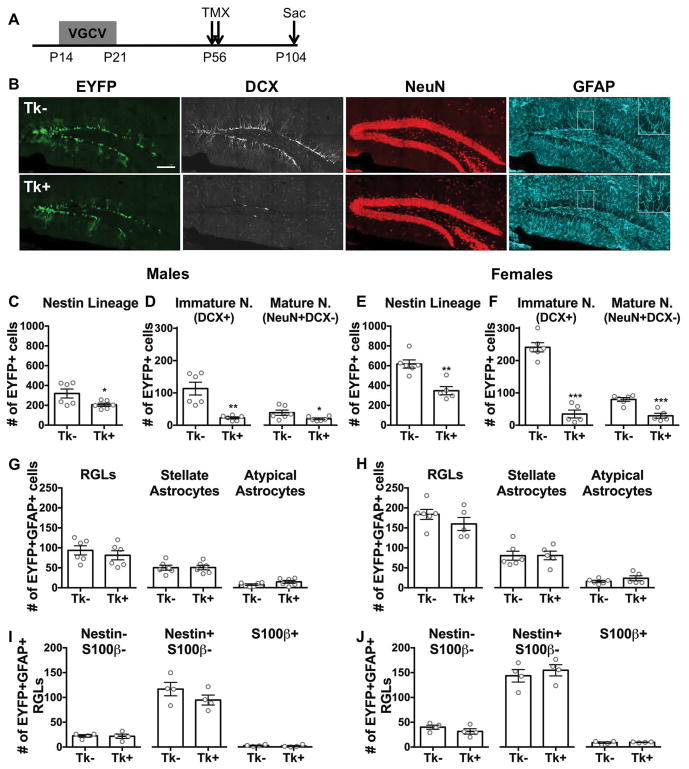
Environmental exposures during early life, but not during adolescence or adulthood, lead to persistent reductions in neurogenesis in the adult hippocampal dentate gyrus (DG). The mechanisms by which early life exposures lead to long-term deficits in neurogenesis remain unclear. Here, we investigated whether targeted ablation of dividing neural stem cells during early life is sufficient to produce long-term decreases in DG neurogenesis. Having previously found that the stem cell lineage is resistant to long-term effects of transient ablation of dividing stem cells during adolescence or adulthood (Kirshenbaum, Lieberman, Briner, Leonardo, & Dranovsky, ), we used a similar pharmacogenetic approach to target dividing neural stem cells for elimination during early life periods sensitive to environmental insults. We then assessed the Nestin stem cell lineage in adulthood. We found that the adult neural stem cell reservoir was depleted following ablation during the first postnatal week, when stem cells were highly proliferative, but not during the third postnatal week, when stem cells were more quiescent. Remarkably, ablating proliferating stem cells during either the first or third postnatal week led to reduced adult neurogenesis out of proportion to the changes in the stem cell pool, indicating a disruption of the stem cell function or niche following stem cell ablation in early life. These results highlight the first three postnatal weeks as a series of sensitive periods during which elimination of dividing stem cells leads to lasting alterations in adult DG neurogenesis and stem cell function. These findings contribute to our understanding of the relationship between DG development and adult neurogenesis, as well as suggest a possible mechanism by which early life experiences may lead to lasting deficits in adult hippocampal neurogenesis.

Keywords: GFAP-Tk; adult neurogenesis; dentate gyrus; early postnatal neurogenesis; hippocampal stem cells.


Figures

Figure 1
Figure 1. Reduction of cell proliferation in the DG of GFAP-Tk mice by VGCV
(A) Experimental timeline of P0–P7 valganciclovir (VGCV) treatment and sacrifice one week after treatment completion in GFAP-Tk+ animals and Tk− controls. (B) The number of CldU+ cells was reduced in Tk+ males and (C) Tk+ females compared to Tk− controls. (D) No difference in the number of Ki67+ cells was detected between Tk− and Tk+ males. (E) The number of Ki67+ cells was reduced in Tk+ versus Tk− females. (F) Experimental timeline of P14–P21 VGCV treatment and sacrifice one week after treatment completion. (G) The number of CldU+ cells was reduced in Tk+ males and (H) females compared to Tk− animals. (I) No difference in the number of Ki67+ cells was detected between Tk− and Tk+ male or (J) female animals. Data are expressed as mean ± SEM. **p<0.01, ***p<0.001
Figure 2
Figure 2. Marker expression and morphology of neurons and astrocytes in the adult Nestin lineage
(A) Representative images of immature neurons co-expressing EYFP and doublecortin (DCX). (B) Representative images of a mature neuron co-expressing EYFP and NeuN. (C) Representative images of a radial glial-like cell (RGL) co-expressing EYFP, GFAP, and Nestin with cell body in the subgranular zone (SGZ) and radial process traversing the granule cell layer. (D) Representative images of stellate astrocytes (SA) co-expressing EYFP and GFAP with multiple similar-sized processes extending from the cell body. Arrowhead points to S100β+ SA. Arrows point to S100β- SAs. (E) Representative images of atypical astrocytes (AA) co-expressing EYFP and GFAP with a single dominant process extending from the cell body, which are in the outer GCL. Arrow points to Nestin+ AA with one process. Arrowhead points to S100β+ AA with one dominant process and multiple minor processes. Scale bar represents 10 μM. Dotted lines indicate SGZ.
Figure 3
Figure 3. Targeting dividing stem cells from P0–P7 leads to depletion of the DG stem cell pool and decreased neurogenesis in adulthood
(A) Experimental timeline of P0–P7 VGCV treatment and tamoxifen (TMX) administration in GFAP-Tk/Nestin-CreERT2 mice. (B) Representative images of EYFP, DCX, NeuN, and GFAP staining in P0–P7 VGCV treated Tk− and Tk+ animals. (C) P0–P7 VGCV led to fewer EYFP+ cells in Tk+ versus Tk− males. (D) P0–P7 VGCV reduced the number of DCX+ immature and NeuN+DCX− mature neurons within the Nestin lineage of Tk+ compared to Tk− males. (E) P0–P7 VGCV led to fewer EYFP+ cells in Tk+ versus Tk− females. (F) P0–P7 VGCV reduced the number of DCX+ immature and NeuN+DCX− mature neurons within the Nestin lineage of Tk+ compared to Tk− females. (G) P0–P7 VGCV decreased the number of RGLs and increased the number of atypical astrocytes, but did not affect the number of stellate astrocytes within the Nestin lineage of Tk+ males and (H) females compared to Tk− animals. (I) P0–P7 VGCV led to fewer Nestin-S100β- and Nestin+S100β- RGLs, but did not change the number of S100β+ (including both Nestin-S100β+ and Nestin+S100β+) RGLs within the Nestin lineage of Tk+ versus Tk− males. (J) P0–P7 VGCV led to fewer Nestin-S100β-, Nestin+S100β-, and S100β+ RGLs within the Nestin lineage of Tk+ versus Tk− females. Scale bar represents 150 μM. GFAP inset is at 2× magnification. Data are expressed as mean ± SEM. *p≤0.05, **p<0.01, ***p<0.001
Figure 4
Figure 4. Targeting dividing stem cells from P14–P21 leads to decreased DG neurogenesis, but does not deplete the stem cell pool in adulthood
(A) Experimental timeline of P14–P21 VGCV treatment and TMX administration in GFAP-Tk/Nestin-CreERT2 mice. (B) Representative images of EYFP, DCX, NeuN, and GFAP staining in P14–P21 VGCV treated Tk− and Tk+ animals. (C) P14–P21 VGCV led to fewer EYFP+ cells in Tk+ versus Tk− males. (D) P14–P21 VGCV reduced the number of DCX+ immature and NeuN+DCX− mature neurons within the Nestin lineage of Tk+ compared to Tk− males. (E) P14–P21 VGCV led to fewer EYFP+ cells in Tk+ versus Tk− females. (F) P14–P21 VGCV reduced the number of DCX+ immature and NeuN+DCX− mature neurons within the Nestin lineage of Tk+ compared to Tk− females. (G) P14–P21 VGCV did not affect the number of RGLs, stellate astrocytes, or atypical astrocytes in the lineage of Tk+ males and (H) females compared to Tk− animals. (I) P14–P21 VGCV did not change the number of Nestin-S100β-, Nestin+S100β- or S100β+ RGLs in the Nestin lineage of Tk+ males and (J) females compared to Tk− animals. Scale bar represents 150 μM. GFAP inset is at 2× magnification. Data are expressed as mean ± SEM. *p≤0.05, **p<0.01, ***p<0.001
Figure 5
Figure 5. More DG stem cells are dividing during the first versus third postnatal week
(A) Experimental timeline of sacrifice of Nestin-Kusabira Orange (KOr) animals at P7 or P21. (B) Representative images of KOr+, MCM2+, and GFAP+ cells in P7 and P21 mice. Arrowheads point to cells expressing all three markers. (C) A larger percentage of KOr+GFAP+ RGLs express the cell division marker MCM2 at P7 compared to at P21 in males and females. Scale bars represent 100 μM for low magnification and 20 μM for high magnification images. Data are expressed as mean ± SEM. ***p<0.001

References

    1. Aisa B, Elizalde N, Tordera R, Lasheras B, Del Rio J, Ramirez MJ. Effects of neonatal stress on markers of synaptic plasticity in the hippocampus: implications for spatial memory. Hippocampus. 2009;19(12):1222–31. - PubMed
    1. Altman J, Bayer SA. Migration and distribution of two populations of hippocampal granule cell precursors during the perinatal and postnatal periods. J Comp Neurol. 1990;301(3):365–81. - PubMed
    1. Alvarez-Buylla A, Lim DA. For the long run: maintaining germinal niches in the adult brain. Neuron. 2004;41(5):683–6. - PubMed
    1. Angevine JB., Jr Time of neuron origin in the hippocampal region. An autoradiographic study in the mouse. Exp Neurol Suppl. 1965;(Suppl 2):1–70. - PubMed
    1. Baek SB, Bahn G, Moon SJ, Lee J, Kim KH, Ko IG, Kim SE, Sung YH, Kim BK, Kim TS, et al. The phosphodiesterase type-5 inhibitor, tadalafil, improves depressive symptoms, ameliorates memory impairment, as well as suppresses apoptosis and enhances cell proliferation in the hippocampus of maternal-separated rat pups. Neurosci Lett. 2011;488(1):26–30. - PubMed

Publication types

MeSH terms

LinkOut - more resources