. 2018 Jun 22;293(25):9724-9735.
doi: 10.1074/jbc.RA118.002248. Epub 2018 May 9.

Human DHEA sulfation requires direct interaction between PAPS synthase 2 and DHEA sulfotransferase SULT2A1

Affiliations

Human DHEA sulfation requires direct interaction between PAPS synthase 2 and DHEA sulfotransferase SULT2A1

Jonathan W Mueller et al. J Biol Chem. .

Abstract

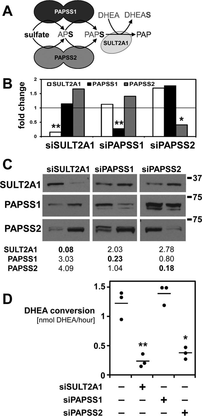
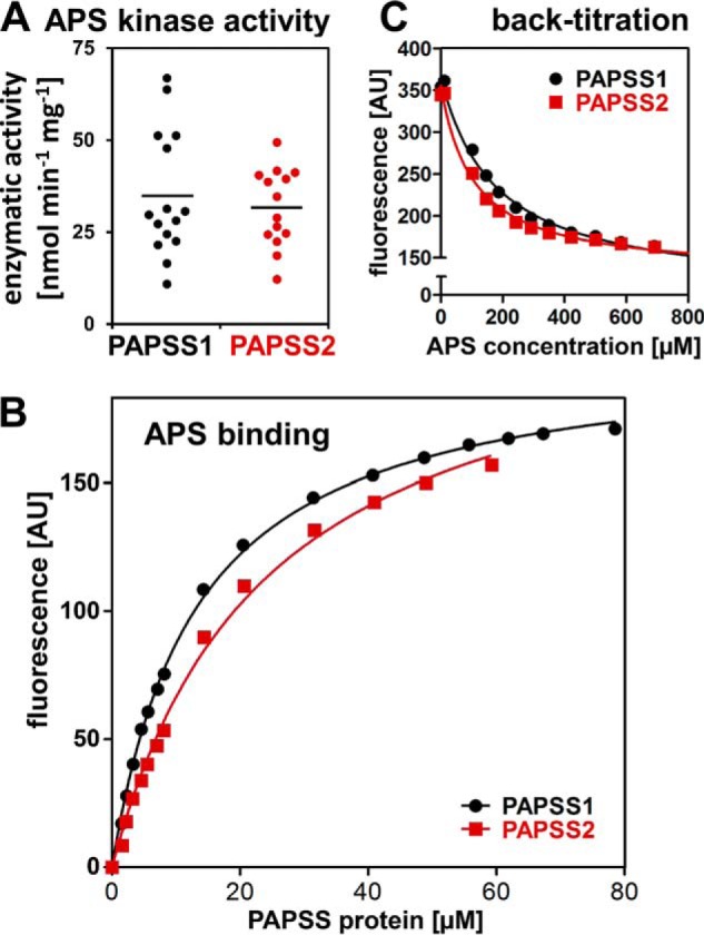
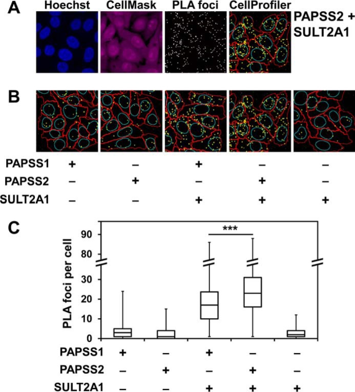
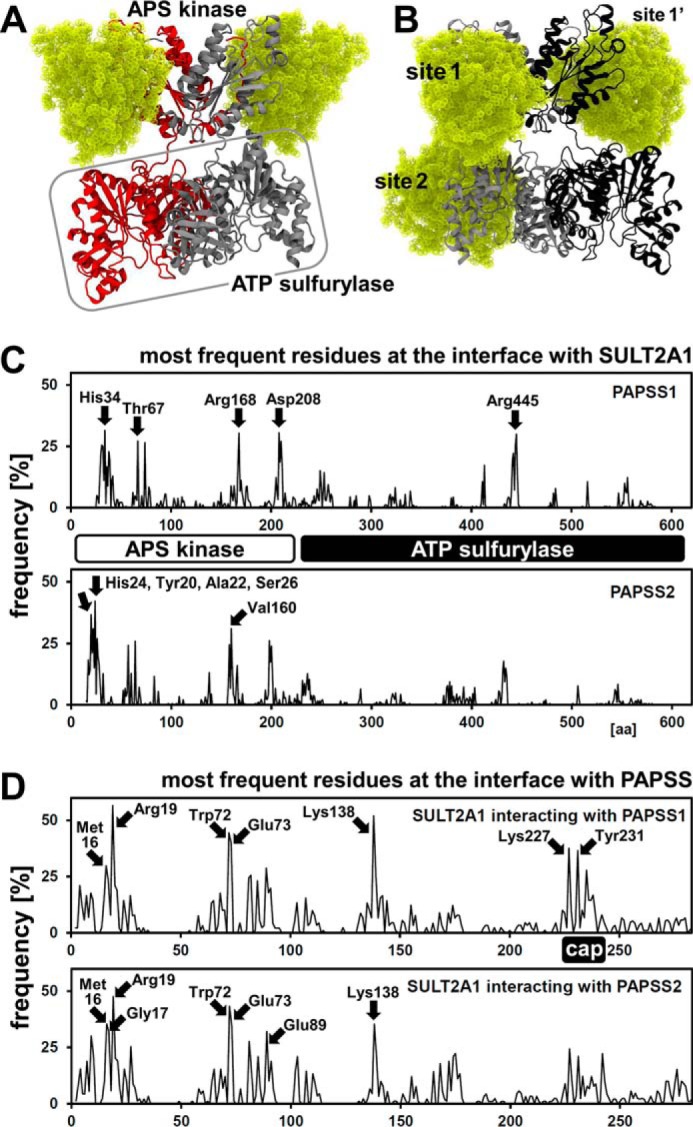
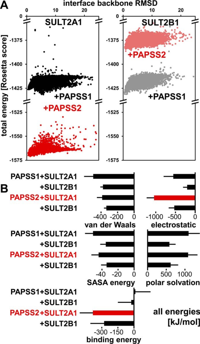
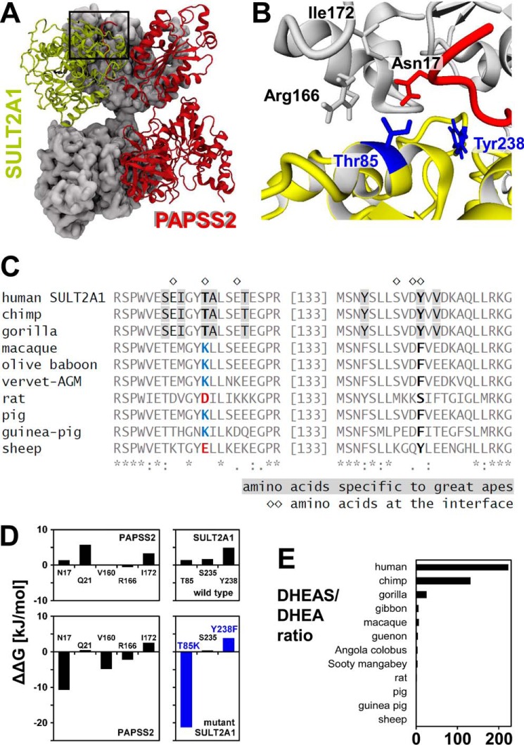
The high-energy sulfate donor 3'-phosphoadenosine-5'-phosphosulfate (PAPS), generated by human PAPS synthase isoforms PAPSS1 and PAPSS2, is required for all human sulfation pathways. Sulfotransferase SULT2A1 uses PAPS for sulfation of the androgen precursor dehydroepiandrosterone (DHEA), thereby reducing downstream activation of DHEA to active androgens. Human PAPSS2 mutations manifest with undetectable DHEA sulfate, androgen excess, and metabolic disease, suggesting that ubiquitous PAPSS1 cannot compensate for deficient PAPSS2 in supporting DHEA sulfation. In knockdown studies in human adrenocortical NCI-H295R1 cells, we found that PAPSS2, but not PAPSS1, is required for efficient DHEA sulfation. Specific APS kinase activity, the rate-limiting step in PAPS biosynthesis, did not differ between PAPSS1 and PAPSS2. Co-expression of cytoplasmic SULT2A1 with a cytoplasmic PAPSS2 variant supported DHEA sulfation more efficiently than co-expression with nuclear PAPSS2 or nuclear/cytosolic PAPSS1. Proximity ligation assays revealed protein-protein interactions between SULT2A1 and PAPSS2 and, to a lesser extent, PAPSS1. Molecular docking studies showed a putative binding site for SULT2A1 within the PAPSS2 APS kinase domain. Energy-dependent scoring of docking solutions identified the interaction as specific for the PAPSS2 and SULT2A1 isoforms. These findings elucidate the mechanistic basis for the selective requirement for PAPSS2 in human DHEA sulfation.

Keywords: DHEAS; PAPS synthase; dehydroepiandrosterone; enzyme kinetics; molecular docking; protein–protein interaction; steroid hormone; steroid sulfation pathway; sulfotransferase.


Conflict of interest statement

The authors declare that they have no conflicts of interest with the contents of this article

Figures

Figure 1.
Figure 1.
Knockdown of components of the DHEA sulfation pathway. A, schematic representation of the DHEA sulfation pathway. Activated sulfate in the form of PAPS is produced by either PAPSS1 or PAPSS2 and then used by the sulfotransferase SULT2A1 to convert DHEA to DHEAS. B and C, siRNA-mediated knockdown of SULT2A1, PAPSS1, or PAPSS2 in adrenocortical NCI-H295R1 cells was verified by real-time PCR and Western blotting. A scrambled oligonucleotide served as control (ctrl). Real-time PCR data normalized to 18S rRNA, fold-change relative to that control. Densitometric quantification of Western blots revealed knockdown efficiencies of up to 90% on the protein level. Double bands were interpreted as degradation products and jointly analyzed. D, DHEA sulfation was assayed for all knockdowns mentioned above, revealing functional differences between PAPSS1 and PAPSS2 for DHEA sulfation by sulfotransferase SULT2A1. Three biological replicates and their average are shown; each dot consists of at least three technical replicates. Normally distributed data were analyzed by one-way ANOVA (p value < 0.001) and post-hoc Bonferroni tests (*, p < 0.05; **, p < 0.01) relative to the control.
Figure 2.
Figure 2.
APS kinase activity and APS binding properties of human PAPS synthases. A, APS kinase activity was measured in a coupled enzymatic assay where ADP production is linked to NADH consumption via pyruvate kinase and lactate dehydrogenase. 15 individual velocity measurements from two different batches are shown. Please refer to Table 1 for the averaged specific activity. B, APS binding studies where fluorescently labeled APS (1 μm mant-APS) was titrated with increasing concentrations of PAPS synthase protein. Data were fitted assuming one binding site. C, back-titration of 1 μm mant-APS and 50 μm PAPSS protein with increasing concentrations of APS. As mant-APS can be displaced by APS, the fluorescent mant moiety did not interfere with binding to the protein.
Figure 3.
Figure 3.
Cytoplasmic PAPSS2 best supports cytoplasmic SULT2A1 activity. A, HEK293 cells were transfected with cytoplasmic sulfotransferase SULT2A1 as well as cytoplasmic (PAPSS1 K9A,K10A and PAPSS2 K6A,K8A) or nuclear protein variants of PAPS synthases (PAPSS1 R111A,R112A and PAPSS2 R101A,R102A) as EGFP fusion proteins. The different variants for PAPSS1 are shown exemplarily; their localization was as described before (18). ×600 magnification. B, DHEA sulfation was assayed for these different PAPS synthase variants. Each point represents the average from triplicate measurements. Normally distributed data were analyzed by one-way ANOVA (p value < 0.001) and post-hoc Bonferroni tests (*, p < 0.05; **, p < 0.01) relative to the control.
Figure 4.
Figure 4.
A physical interaction of PAPSS2 and SULT2A1 detected by proximity ligation assays. A, representative images of the proximity ligation assay between PAPSS2 and SULT2A1 as well as subsequent analysis with CellProfiler. Endogenous PAPS synthases and SULT2A1 were detected by mouse monoclonal antibodies for PAPSS1 or PAPSS2 and a rabbit SULT2A1 polyclonal antibody in a HepG2 cell line. PLA analysis, including automated cell and nucleus recognition and foci counting was carried out using CellProfiler software. Cell nuclei were stained with Hoechst 33342 (blue); CellMask staining is shown in magenta. PLA foci are shown in white in the single channel picture. In the output image of CellProfiler analysis edges of nuclei are represented in cyan, cell boarders in red, and PLA foci in yellow. B, CellProfiler results for all other combinations. Negative controls were generated using only one primary antibody at a time. ×600 magnification for A and B. C, box-and-whisker analysis of the PLA foci number per cell from at least 400 cells pooled from three independent experiments. Data were found to be not normally distributed; hence, one-way ANOVA (p value < 0.001) and post hoc Bonferroni tests (***, p < 0.001) were performed after data were square root transformed.
Figure 5.
Figure 5.
SULT2A1 docks to the APS kinase domain of PAPSS2. ClusPro computational docking of three different SULT2A1 crystal structures (PDB codes 1EFH, 3F3Y, and 4IFB) to structural models of PAPSS1 and PAPSS2. A, PAPSS2–SULT2A1-docked complexes are shown. PAPSS2 APS kinase is labeled; ATP sulfurylase is labeled and boxed. The two PAPSS2 dimeric subunits are gray and red. Two SULT2A1 molecules are depicted in yellow, ball representation; contacting PAPSS2 at its APS kinase domain, sites 1 and 1′. B, corresponding representation of PAPSS1–SULT2A1 docking experiments. In addition to sites 1 and 1′, SULT2A1 contacts PAPSS1 also at the ATP sulfurylase domain. Color coding as in A, except the two PAPSS1 dimeric subunits, which are gray and black. C and D, statistical analysis of all ClusPro docking experiments, looking from the PAPS synthase side (C) and from the SULT2A1 side (D). Frequency of individual residues within 3 Å of the other protein was analyzed for PDB 1EFH, 3F3Y, and 4IFB structures separately (30 dockings each) and then averaged. Note the higher number of frequent protein contacts within the APS kinase domain of PAPSS2, compared with the one from PAPSS1. SULT2A1 contacted PAPS synthases mainly via its isoform-specific substrate binding loops; one of these is regarded as “cap.”
Figure 6.
Figure 6.
The PAPSS2–SULT2A1 interaction may be isoform-specific. A, SULT2B1 was selected as homologous sulfotransferase to analyze specificity of the novel PAPSS2–sulfotransferase interaction. PAPS synthase–sulfurylase docking was refined using RosettaDock. At least 10,000 docking experiments are shown where the docking score was correlated with the interface r.m.s. deviation value compared with the average complex. B, best solutions from Rosetta were subjected to MD simulations (3 × 20 ns, see Fig. S2 for averaged traces); MM-PBSA energies were derived therefrom, expressed as average ± S.D. from three independent calculations.
Figure 7.
Figure 7.
A PAPSS2–SULT2A1 protein interaction facilitates DHEA sulfation. A and B, molecular representation of a PAPSS2–SULT2A1 complex averaged over 15 ns of MD time. The dimeric PAPSS2 subunit proximal to SULT2A1 is depicted in gray color and with molecular surface representation; the distant PAPSS2 subunit in red color and ribbon representation. SULT2A1 is drawn in yellow. Please note the composite nature of the PAPSS2-binding site. Amino acids on the interface are shown in stick representation of the side chains and labeled accordingly. C, all SULT2A1 amino acids on the interface with PAPSS2 were highlighted in an alignment of diverse mammalian SULT2A1 protein sequences. The only two interface amino acids that were specific to great apes are Thr85 and Tyr238 (depicted in blue in B). D, the PAPSS2–SULT2A1 interface was analyzed using Rosetta-based alanine scanning (27). Furthermore, the two great ape-specific amino acids were mutated to their nonhominoid counterparts. T85K resulted in a dramatic loss of stability of the complex. E, the hominid-specific PAPSS2–SULT2A1 complex coincides with a higher DHEAS/DHEA ratio in gorilla, chimpanzee, and human. DHEAS/DHEA ratios are derived from Refs. and .

References

    1. Strott C. A. (2002) Sulfonation and molecular action. Endocr. Rev. 23, 703–732 10.1210/er.2001-0040 - DOI - PubMed
    1. Mueller J. W., Gilligan L. C., Idkowiak J., Arlt W., and Foster P. A. (2015) The regulation of steroid action by sulfation and desulfation. Endocr. Rev. 36, 526–563 10.1210/er.2015-1036 - DOI - PMC - PubMed
    1. Herrero J., Muffato M., Beal K., Fitzgerald S., Gordon L., Pignatelli M., Vilella A. J., Searle S. M., Amode R., Brent S., Spooner W., Kulesha E., Yates A., and Flicek P. (2016) Ensembl comparative genomics resources. Database (Oxford) 2016, pii:baw053 - PMC - PubMed
    1. van den Boom J., Heider D., Martin S. R., Pastore A., and Mueller J. W. (2012) 3′-Phosphoadenosine 5′-phosphosulfate (PAPS) synthases, naturally fragile enzymes specifically stabilized by nucleotide binding. J. Biol. Chem. 287, 17645–17655 10.1074/jbc.M111.325498 - DOI - PMC - PubMed
    1. Kauffman F. C. (2004) Sulfonation in pharmacology and toxicology. Drug Metab. Rev. 36, 823–843 10.1081/DMR-200033496 - DOI - PubMed

Publication types

MeSH terms

LinkOut - more resources