. 2018 Jun 4;150(6):851-862.
doi: 10.1085/jgp.201711968. Epub 2018 May 9.

Histidine168 is crucial for ΔpH-dependent gating of the human voltage-gated proton channel, hHV1

Affiliations

Histidine168 is crucial for ΔpH-dependent gating of the human voltage-gated proton channel, hHV1

Vladimir V Cherny et al. J Gen Physiol. .

Abstract

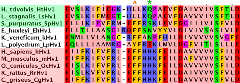
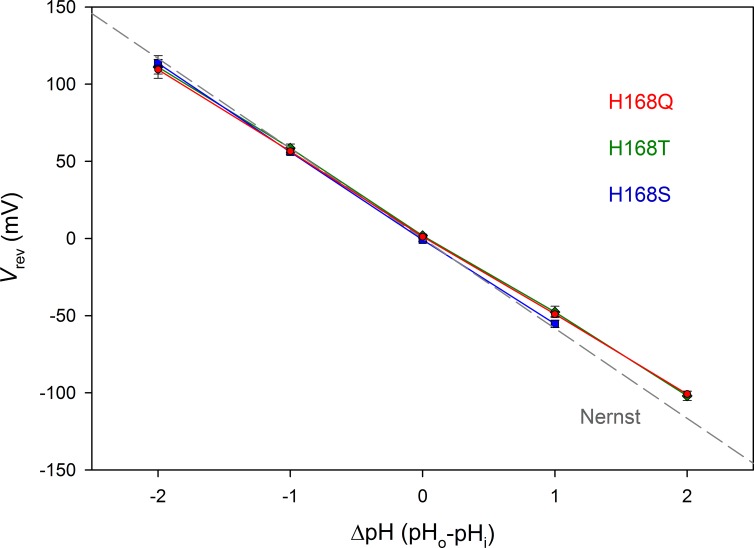
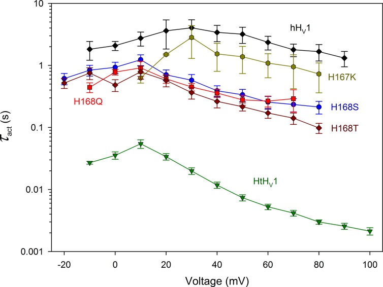
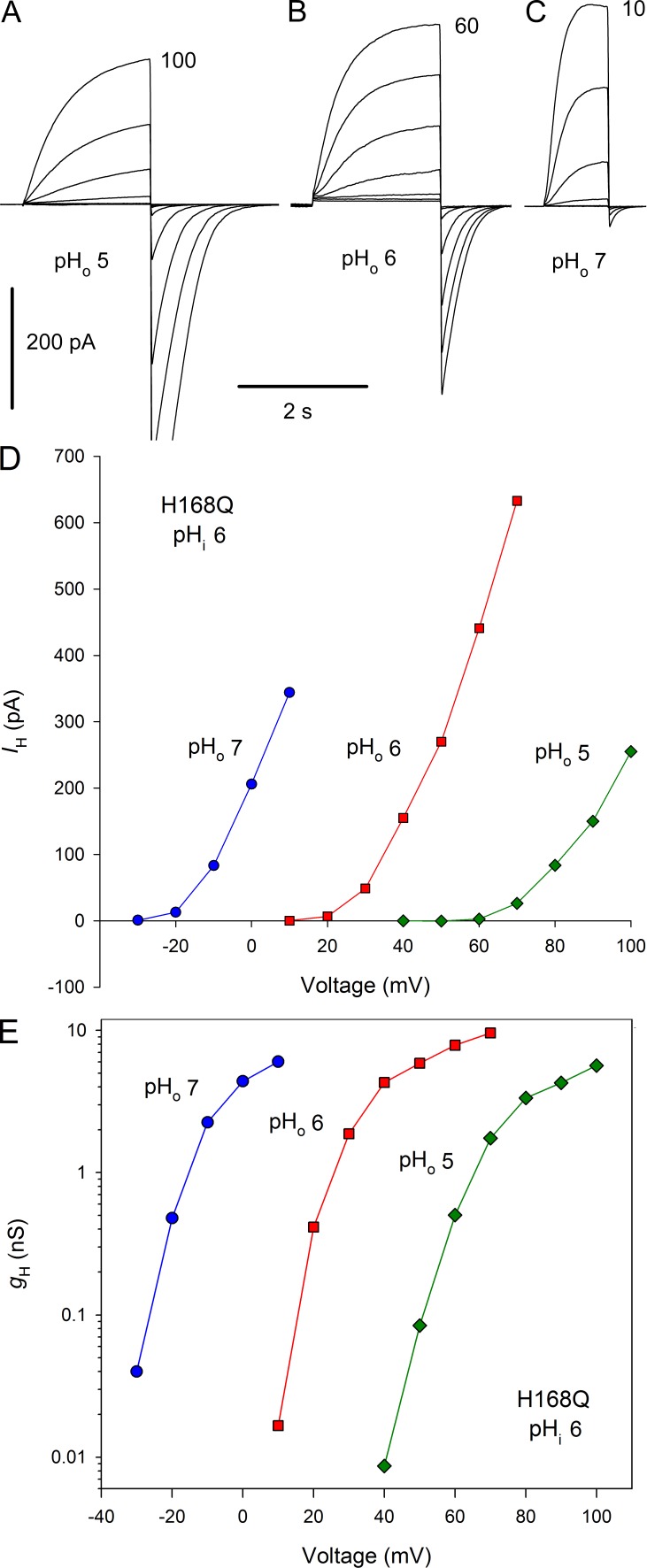
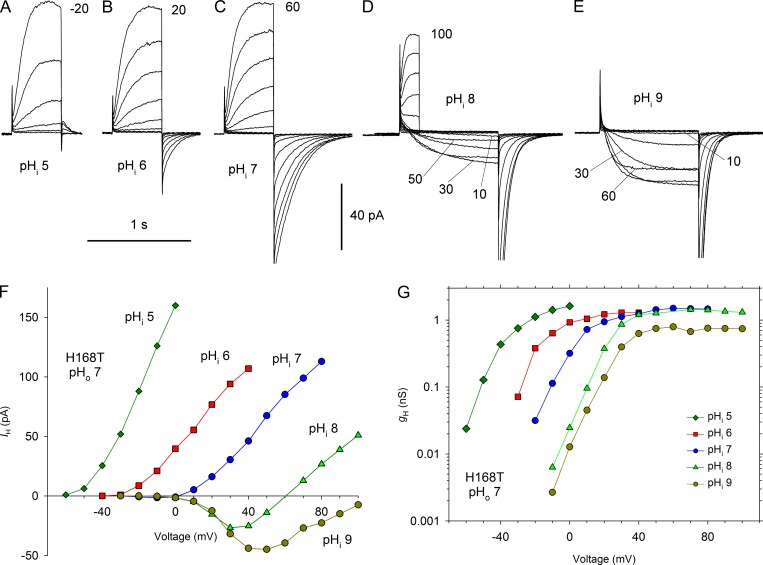
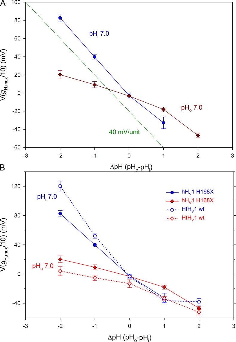
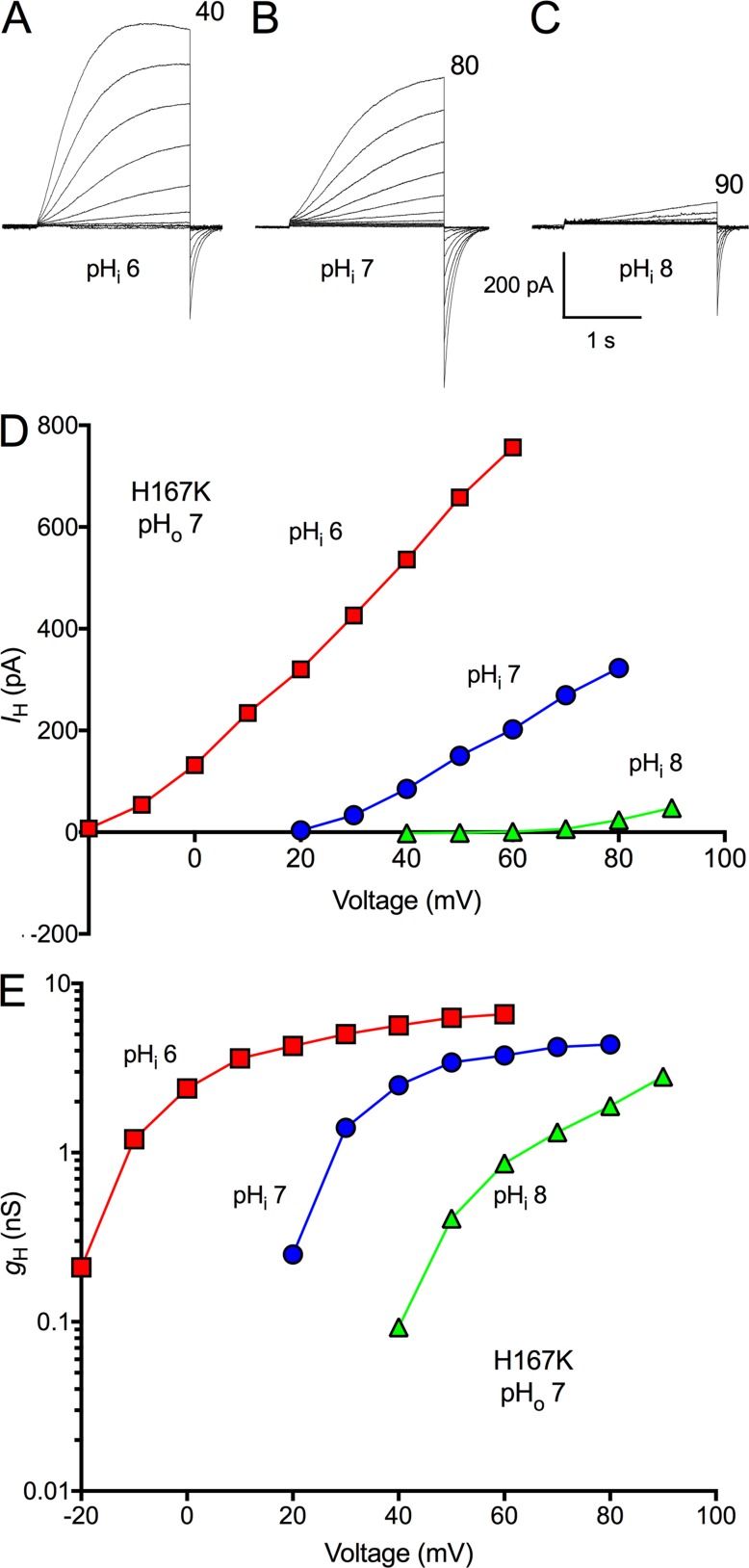
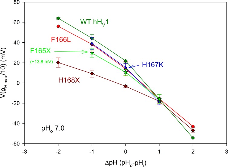
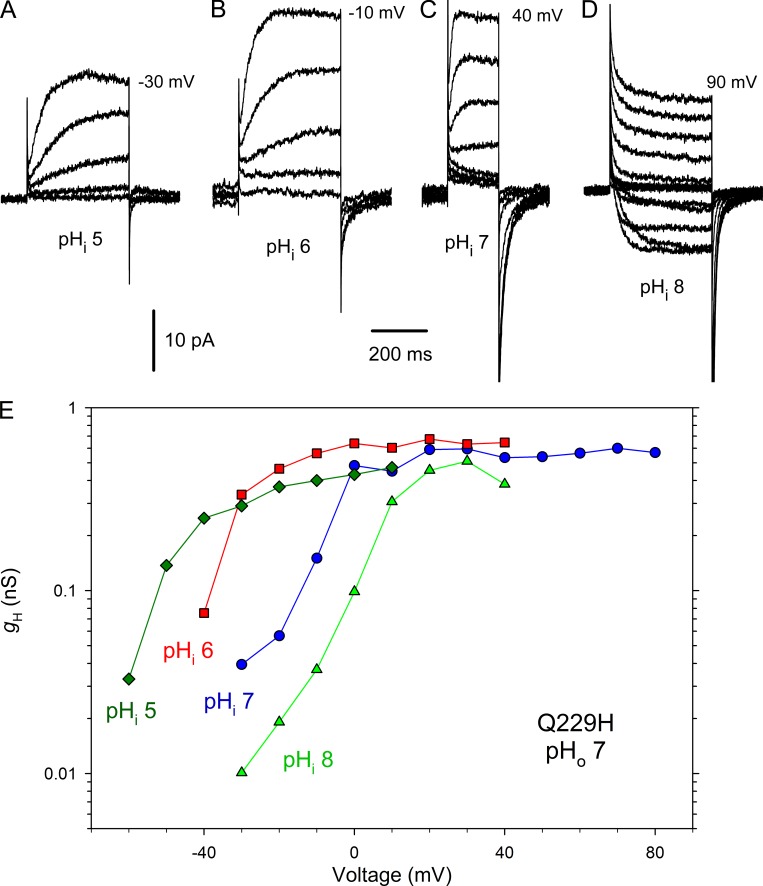
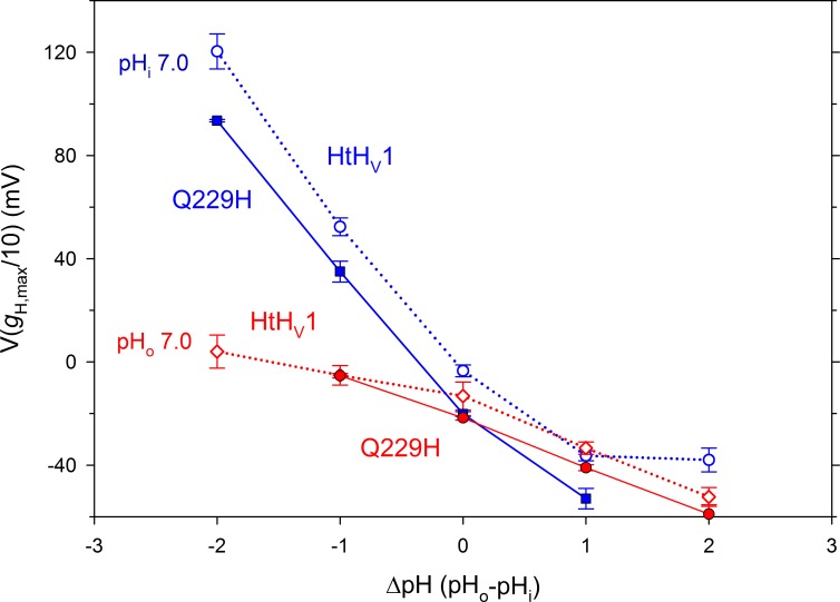
We recently identified a voltage-gated proton channel gene in the snail Helisoma trivolvis, HtHV1, and determined its electrophysiological properties. Consistent with early studies of proton currents in snail neurons, HtHV1 opens rapidly, but it unexpectedly exhibits uniquely defective sensitivity to intracellular pH (pHi). The H+ conductance (gH)-V relationship in the voltage-gated proton channel (HV1) from other species shifts 40 mV when either pHi or pHo (extracellular pH) is changed by 1 unit. This property, called ΔpH-dependent gating, is crucial to the functions of HV1 in many species and in numerous human tissues. The HtHV1 channel exhibits normal pHo dependence but anomalously weak pHi dependence. In this study, we show that a single point mutation in human hHV1-changing His168 to Gln168, the corresponding residue in HtHV1-compromises the pHi dependence of gating in the human channel so that it recapitulates the HtHV1 response. This location was previously identified as a contributor to the rapid gating kinetics of HV1 in Strongylocentrotus purpuratus His168 mutation in human HV1 accelerates activation but accounts for only a fraction of the species difference. H168Q, H168S, or H168T mutants exhibit normal pHo dependence, but changing pHi shifts the gH-V relationship on average by <20 mV/unit. Thus, His168 is critical to pHi sensing in hHV1. His168, located at the inner end of the pore on the S3 transmembrane helix, is the first residue identified in HV1 that significantly impairs pH sensing when mutated. Because pHo dependence remains intact, the selective erosion of pHi dependence supports the idea that there are distinct internal and external pH sensors. Although His168 may itself be a pHi sensor, the converse mutation, Q229H, does not normalize the pHi sensitivity of the HtHV1 channel. We hypothesize that the imidazole group of His168 interacts with nearby Phe165 or other parts of hHV1 to transduce pHi into shifts of voltage-dependent gating.


Figures

Figure 1.
Figure 1.
Alignment of HV1 S2–S3 linker and nearby regions (E153–D185 in hHV1). HV1 from several species were characterized as activating rapidly (green background: HtHV1, LsHV1, and SpHV1) or slowly (pink background: hHV1, mHV1, OcHV1, RrHV1, and CgHV1) based on available electrophysiological data. Unshaded species exhibit intermediate kinetics. In hHV1 numbering, the asterisk (*) indicates the position corresponding to the throttle histidine H168; the caret (^) indicates the position corresponding to F165. The S2–S3 linker in hHV1 encompasses K157–F165 (Li et al., 2015).
Figure 2.
Figure 2.
His168 mutants are proton selective. Mean ± SEM values for the reversal potential (Vrev) are plotted for 12 H168Q, 9 H168T, and 6 H168S cells or patches. The linear regression slope for each mutant is −52.6, −53.2, and −56.3 mV/unit change in ΔpH, respectively. For comparison, the gray dashed line shows the Nernst potential, EH, expected for perfect H+ selectivity.
Figure 3.
Figure 3.
The throttle histidine explains only a fraction of the rapid activation of HtHV1. Activation time constants (τact) were determined by single exponential fits to currents in WT HtHV1, WT hHV1, and in four hHV1 mutants, as indicated, all at pHo 7 and pHi 7. Mean ± SEM is plotted for 3–12 WT hHV1, 2–3 H167K, 3–6 H168S, 3–9 H168Q, 3–6 H168T, and 7–17 WT HtHV1. At all voltages from −10 to +80 mV, the WT hHV1 value is significantly larger than that of H168T, H168S, and H168Q combined and larger than HtHV1 (P < 0.01). The WT hHV1 data were taken from Cherny et al. (2015) and the HtHV1 data from Thomas et al. (2018).
Figure 4.
Figure 4.
Gating of His168 mutants has normal pHo dependence. (A–C) Families of currents in a cell expressing H168Q with pHi 6 and pHo 5, 6, and 7, with pulses in 10-mV increments up to the voltage indicated. Holding potential, Vhold was −40 mV (A and B) or −60 mV (C). (D and E) Proton current–voltage curves (D) and gH-V relationships (E) from the families in A–C exhibit a normal 40-mV shift/unit change in pHo.
Figure 5.
Figure 5.
Mutation of His168 in hHV1 weakens the pHi dependence of gating. (A–E) Families of currents at several pHi values in an inside-out patch from a cell transfected with H168T, with pHo 7 in the pipette. Pulses were applied in 10-mV increments up to the voltage indicated from Vhold = −80 mV (A), −60 mV (B), or −40 mV (C–E). (D) Shorter pulses were applied to 60 mV and above, and the tail currents have been removed. (E) Pulses above 60 mV were omitted for clarity. (F) Current–voltage curves. (G) gH-V relationships from this experiment.
Figure 6.
Figure 6.
The His168 mutation recapitulates the anomalous ΔpH dependence of HtHV1. (A) The effect of pHo and pHi on the position of the gH-V relationship in the three H168x mutants combined (mean ± SEM) is plotted. The position of the gH-V relationship was defined in terms of V(gH,max/10). The dashed green line shows a slope of 40 mV/unit for reference. The dependence of these mutants on pHo is normal, whereas their pHi response is greatly attenuated. Linear regression on all points (ignoring the obvious nonlinearity) gives a slope of 38.9 mV/unit change in pHo and 16.1 mV/unit change in pHi. (B) The same data are replotted (shaded symbols) along with the analogous measurements for HtHV1 (open symbols) taken from Fig. 9 of the companion article (Thomas et al., 2018). In whole-cell measurements, pHo was varied with pHi 7 (blue symbols). When pHi was varied by using inside-out patches with pHo 7, there was very little shift of the gH-V relationship (red symbols). Numbers of cells for increasing ΔpH in H168X mutants for pHi 7 are 3, 10, 11, and 5 and for pHo 7 are 6, 10, 14, 11, and 4.
Figure 7.
Figure 7.
The H167K mutant of hHV1 has normal pHi dependence. (A–C) Families of currents are shown in an inside-out patch with pHo 7 and pHi 6, 7, or 8, as labeled. Pulses were applied in 10-mV increments up to the voltage indicated, from Vhold = −60 mV (A) or -40 mV (B and C). (D and E) Current–voltage curves (D) and gH-V relationships from this patch are illustrated (E). At pHi 8, gH was estimated from tail current amplitudes and scaled according to gH calculated from the outward current at 90 mV.
Figure 8.
Figure 8.
The F166L and H167K mutations do not impair pHi sensing of hHV1. The pHi responses of the H168X mutants are replotted from Fig. 6. The pHi responses of WT hHV1, the F166L mutant of hHV1, and the H167K mutant of hHV1 are indistinguishable from each other. The F165X data are from three F165A and three F165H patches combined, and 13.8 mV has been added to all values to facilitate comparison with WT. All data (mean ± SEM) come from inside-out patches studied at pHo 7, with n = 2–9 (H168X), 1–4 (F166L), 3 (H167K), and 6 (F165X).
Figure 9.
Figure 9.
The Q229H mutation does not restore human-like activation kinetics or pHi sensitivity to HtHV1. (A–D) Families of currents in an inside-out patch with pHo 7 in the pipette solution in 10-mV increments up to the voltage shown from Vhold = −80, −60, −40, and −40 mV, at pHi 5, 6, 7, and 8, respectively. (E) Currents from this patch converted to gH produce gH-V relationships that shift much less than 40 mV/unit. Corresponding measurements in WT HtHV1 are plotted in Fig. 5 G.
Figure 10.
Figure 10.
The Q229H mutation does not restore human-like pHi sensitivity to HtHV1. The dependence of the position of the gH-V relationship on both pHo and pHi is similar in WT HtHV1 (open symbols and dotted lines) and in the Q229H mutant (shaded symbols and solid lines). The WT data are replotted from Fig. 6 B. Mean ± SEM are shown for n = 2–5 cells with pHi 7 and three inside-out patches with pHo 7.

Comment in

References

    1. Bennett A.L., and Ramsey I.S.. 2017. CrossTalk opposing view: proton transfer in Hv1 utilizes a water wire, and does not require transient protonation of a conserved aspartate in the S1 transmembrane helix. J. Physiol. 595:6797–6799. 10.1113/JP274553 - DOI - PMC - PubMed
    1. Berger T.K., Fußhöller D.M., Goodwin N., Bönigk W., Müller A., Dokani Khesroshahi N., Brenker C., Wachten D., Krause E., Kaupp U.B., and Strünker T.. 2017. Post-translational cleavage of Hv1 in human sperm tunes pH- and voltage-dependent gating. J. Physiol. 595:1533–1546. 10.1113/JP273189 - DOI - PMC - PubMed
    1. Bernheim L., Krause R.M., Baroffio A., Hamann M., Kaelin A., and Bader C.R.. 1993. A voltage-dependent proton current in cultured human skeletal muscle myotubes. J. Physiol. 470:313–333. 10.1113/jphysiol.1993.sp019860 - DOI - PMC - PubMed
    1. Byerly L., Meech R., and Moody W. Jr. 1984. Rapidly activating hydrogen ion currents in perfused neurones of the snail, Lymnaea stagnalis. J. Physiol. 351:199–216. 10.1113/jphysiol.1984.sp015241 - DOI - PMC - PubMed
    1. Chakraborty A., Mandloi S., Lanczycki C.J., Panchenko A.R., and Chakrabarti S.. 2012. SPEER-SERVER: a web server for prediction of protein specificity determining sites. Nucleic Acids Res. W242-W248 10.1093/nar/gks559 - DOI - PMC - PubMed

Publication types