. 2018 Jun 29;92(14):e00396-18.
doi: 10.1128/JVI.00396-18. Print 2018 Jul 15.

Influenza A Virus Infection Triggers Pyroptosis and Apoptosis of Respiratory Epithelial Cells through the Type I Interferon Signaling Pathway in a Mutually Exclusive Manner

Affiliations

Influenza A Virus Infection Triggers Pyroptosis and Apoptosis of Respiratory Epithelial Cells through the Type I Interferon Signaling Pathway in a Mutually Exclusive Manner

SangJoon Lee et al. J Virol. .

Abstract

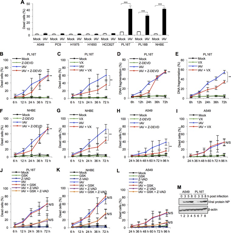
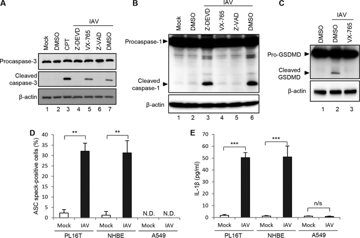
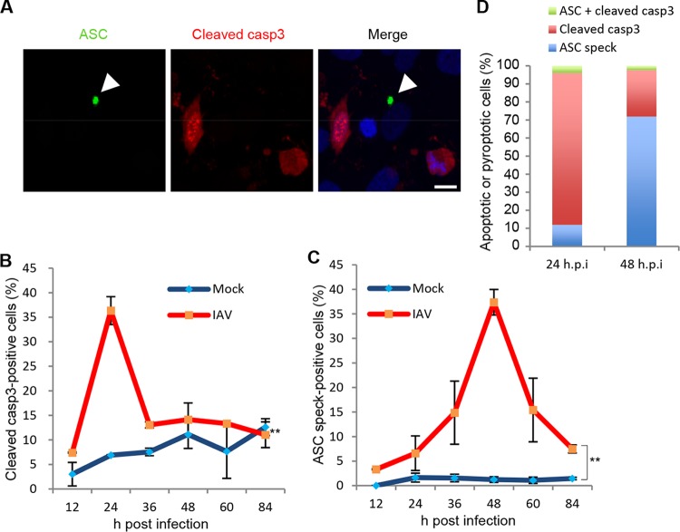
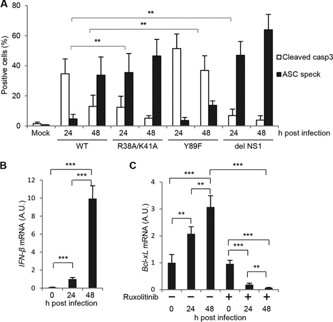
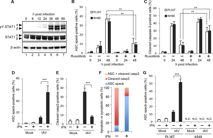
Respiratory epithelial cell death by influenza virus infection is responsible for the induction of inflammatory responses, but the exact cell death mechanism is not understood. Here we showed that influenza virus infection induces apoptosis and pyroptosis in normal or precancerous human bronchial epithelial cells. Apoptosis was induced only in malignant tumor cells infected with influenza virus. In human precancerous respiratory epithelial cells (PL16T), the number of apoptotic cells increased at early phases of infection, but pyroptotic cells were observed at late phases of infection. These findings suggest that apoptosis is induced at early phases of infection but the cell death pathway is shifted to pyroptosis at late phases of infection. We also found that the type I interferon (IFN)-mediated JAK-STAT signaling pathway promotes the switch from apoptosis to pyroptosis by inhibiting apoptosis possibly through the induced expression of the Bcl-xL anti-apoptotic gene. Further, the inhibition of JAK-STAT signaling repressed pyroptosis but enhanced apoptosis in infected PL16T cells. Collectively, we propose that type I IFN signaling pathway triggers pyroptosis but not apoptosis in the respiratory epithelial cells in a mutually exclusive manner to initiate proinflammatory responses against influenza virus infection.IMPORTANCE Respiratory epithelium functions as a sensor of infectious agents to initiate inflammatory responses along with cell death. However, the exact cell death mechanism responsible for inflammatory responses by influenza virus infection is still unclear. We showed that influenza virus infection induced apoptosis and pyroptosis in normal or precancerous human bronchial epithelial cells. Apoptosis was induced at early phases of infection, but the cell death pathway was shifted to pyroptosis at late phases of infection under the regulation of type I IFN signaling to promote proinflammatory cytokine production. Taken together, our results indicate that the type I IFN signaling pathway plays an important role to induce pyroptosis but represses apoptosis in the respiratory epithelial cells to initiate proinflammatory responses against influenza virus infection.

Keywords: apoptosis; pyroptosis; respiratory epithelial cells; type I IFN signaling.


Figures

FIG 1
FIG 1
Influenza virus infection induces pyroptotic cell death in precancerous respiratory epithelial cells. (A) The numbers of dead cells by IAV infection in human malignant respiratory epithelial cells (A549, PC9, H1975, H1650, HCC827), human atypical adenomatous hyperplasia (AAH) respiratory epithelial cells (PL16T), human nonneoplastic respiratory epithelial cells (PL16B), and primary normal human bronchial epithelial cells (NHBE) were determined by trypan blue dye exclusion assays. The data represent averages with standard deviations from three independent experiments (n > 100). ***, P < 0.001 by Student's t test. (B to L) PL16T cells (B, C, D, E, and J), NHBE cells (F, G, and K), and A549 cells (H, I, and L) were infected with IAV at an MOI of 10 in the presence of 10 μM Z-DEVD-FMK (Z-DEVD; caspase-3 inhibitor) (B, D, F, and H), 20 μM VX-765 (VX; caspase-1 inhibitor) (C, E, G, and I), and 3 μM GSK-872 (GSK; RIP3 inhibitor) with 20 μM Z-VAD-FMK (Z-VAD; pan-caspase inhibitor) (J, K, and L). The number of dead cells was measured by trypan blue dye exclusion assays, and the cells with fragmented DNA were quantified by ArrayScan high-content systems. The data represent averages with standard deviations from three independent experiments (n > 100). **, P < 0.01 by two-way analysis of variance (ANOVA); N/S, not significant. (M) At 3, 5, and 8 h postinfection, infected A549 (lanes 1 to 4) and PL16T cells (lanes 5 to 8) were lysed, and the lysates were analyzed by SDS-PAGE followed by Western blotting assays using anti-NP and anti-β-actin antibodies.
FIG 2
FIG 2
IAV infection induces inflammasome assembly and IL-1β secretion in human respiratory epithelial cells. (A) Infected PL16T cells were incubated with either 10 μM Z-DEVD-FMK, 20 μM VX-765, or 20 μM Z-VAD-FMK for 24 h, and then the cell lysates were subjected to Western blot analysis with anti-procaspase-3 and anti-cleaved caspase-3 antibodies. As a positive control, cells were treated with 10 μM camptothecin (CPT) to activate caspase-3. β-Actin was detected as a loading control. DMSO, dimethyl sulfoxide. (B) Infected PL16T cells were incubated with either 10 μM Z-DEVD-FMK, 20 μM VX-765, or 20 μM Z-VAD-FMK for 48 h, and then the cell lysates were subjected to Western blot analysis using anti-caspase-1 antibody. β-Actin was detected as a loading control. (C) Infected PL16T cells were incubated with 20 μM VX-765 for 48 h, and then the cell lysates were subjected to Western blot analysis using anti-GSDMD antibody. β-Actin was detected as a loading control. (D) At 48 h postinfection, uninfected and infected PL16T, NHBE, and A549 cells were subjected to indirect immunofluorescence assays using anti-ASC antibody, and the percentages of ASC speck-positive cells were quantitated. The data represent averages with standard deviations from three independent experiments (n > 100). (E) PL16T, NHBE, and A549 cells were infected with IAV at an MOI of 10. At 72 h postinfection, cell-free supernatants were collected and the concentration of IL-1β was quantified by ELISA. The data represent averages with standard deviations from three independent experiments. **, P < 0.01; ***, P < 0.001 by Student's t test; N.D., not detected; n/s, not significant.
FIG 3
FIG 3
Apoptosis and pyroptosis are independently induced in PL16T cells against IAV infection. (A to D) At 12, 24, 36, 48, 60, and 84 h postinfection, uninfected and infected PL16T cells were subjected to indirect immunofluorescence assays with anti-ASC (green) and anti-cleaved caspase-3 (red) antibodies. A representative result at 24 h postinfection is shown in panel A. Arrowheads indicate ASC inflammasome. Bar, 10 μm. The average numbers of cleaved caspase-3-positive (B) and ASC speck-positive (C) cells and standard deviations obtained from three independent experiments are shown (n > 100). The percentages of cleaved caspase-3-positive (red), ASC speck-positive (blue), and cleaved caspase-3/ASC speck-positive (green) cells were determined at 24 and 48 h postinfection (D). **, P < 0.01 by two-way ANOVA.
FIG 4
FIG 4
Apoptotic and pyroptotic cell death of respiratory epithelial cells induced by NS1 mutant viruses. (A) PL16T cells were infected with either wild-type, R38A/K41A, Y89F, or del NS1 virus. At 24 and 48 h postinfection, the cells were subjected to indirect immunofluorescence assays with anti-ASC and anti-cleaved caspase-3 antibodies. The average numbers of cleaved caspase-3-positive and ASC speck-positive cells and standard deviations obtained from three independent experiments are shown (n > 100). (B) PL16T cells were infected with wild-type IAV. At 24 and 48 h postinfection, cells were collected and the total RNAs were subjected to reverse transcription followed by real-time PCR with primers specific for IFN-β mRNA. The mean values and standard deviations obtained from three independent experiments are shown. (C) PL16T cells were infected with wild-type IAV in the absence or presence of 1 μg/ml ruxolitinib. At 24 and 48 h postinfection, total RNAs were purified and were subjected to reverse transcription followed by real-time PCR with primers specific for Bcl-xL mRNA. The mean values and standard deviations obtained from three independent experiments are shown. **, P < 0.01; ***, P < 0.001 by Student's t test.
FIG 5
FIG 5
Type I IFN triggers pyroptosis but not apoptosis in a mutually exclusive manner. (A) At 0, 6, 12, 24, 36, 48, and 60 h postinfection, infected PL16T cells were lysed, and the cell lysates were subjected to Western blot analysis using anti-phospho-STAT1 (Tyr701) and STAT1. β-Actin was detected as a loading control. (B and C) PL16T (open bars) and NHBE (filled bars) cells were infected with IAV at an MOI of 10 in the absence or presence of 1 μg/ml ruxolitinib. At 24 and 48 h postinfection, PL16T and NHBE cells were subjected to indirect immunofluorescence assays using anti-cleaved caspase-3 and anti-ASC antibodies. The percentage of ASC speck-positive cells (B) and that of cleaved caspase-3-positive cells (C) are shown. The mean values and standard deviations obtained from three independent experiments are shown (n > 100). (D to F) At 6 h postinfection, infected PL16T cells were incubated with or without 1,000 IU/ml IFN-β. At 24 h postinfection, cells were subjected to indirect immunofluorescence assays using anti-ASC and anti-cleaved caspase-3 antibodies. The average numbers of ASC speck-positive (D) and cleaved caspase-3-positive (E) cells and standard deviations obtained from three independent experiments are shown (n > 100). The percentages of ASC speck-positive (blue), cleaved caspase-3-positive (red), and cleaved caspase-3/ASC speck-positive (green) cells were determined (F). (G) At 6 h postinfection, infected PL16T and A549 cells were incubated with or without 1,000 IU/ml IFN-β. At 24 h postinfection, cells were subjected to indirect immunofluorescence assays using anti-ASC antibody. The average numbers of ASC speck-positive cells and standard deviations obtained from three independent experiments are shown (n > 100). **, P < 0.01; ***, P < 0.001 by Student's t test; N.D., not detected.

Similar articles

Cited by

References

    1. Fink SL, Cookson BT. 2005. Apoptosis, pyroptosis, and necrosis: mechanistic description of dead and dying eukaryotic cells. Infect Immun 73:1907–1916. doi:10.1128/IAI.73.4.1907-1916.2005. - DOI - PMC - PubMed
    1. Ashida H, Mimuro H, Ogawa M, Kobayashi T, Sanada T, Kim M, Sasakawa C. 2011. Cell death and infection: a double-edged sword for host and pathogen survival. J Cell Biol 195:931–942. doi:10.1083/jcb.201108081. - DOI - PMC - PubMed
    1. Lamkanfi M, Dixit VM. 2010. Manipulation of host cell death pathways during microbial infections. Cell Host Microbe 8:44–54. doi:10.1016/j.chom.2010.06.007. - DOI - PubMed
    1. Creagh EM. 2014. Caspase crosstalk: integration of apoptotic and innate immune signalling pathways. Trends Immunol 35:631–640. doi:10.1016/j.it.2014.10.004. - DOI - PubMed
    1. Cho YS, Challa S, Moquin D, Genga R, Ray TD, Guildford M, Chan FK. 2009. Phosphorylation-driven assembly of the RIP1-RIP3 complex regulates programmed necrosis and virus-induced inflammation. Cell 137:1112–1123. doi:10.1016/j.cell.2009.05.037. - DOI - PMC - PubMed

Publication types

MeSH terms

Substances