. 2018 Jun 29;92(14):e02222-17.
doi: 10.1128/JVI.02222-17. Print 2018 Jul 15.

Novel Strategy To Adapt Simian-Human Immunodeficiency Virus E1 Carrying env from an RV144 Volunteer to Rhesus Macaques: Coreceptor Switch and Final Recovery of a Pathogenic Virus with Exclusive R5 Tropism

Affiliations

Novel Strategy To Adapt Simian-Human Immunodeficiency Virus E1 Carrying env from an RV144 Volunteer to Rhesus Macaques: Coreceptor Switch and Final Recovery of a Pathogenic Virus with Exclusive R5 Tropism

Hanna B Scinto et al. J Virol. .

Abstract

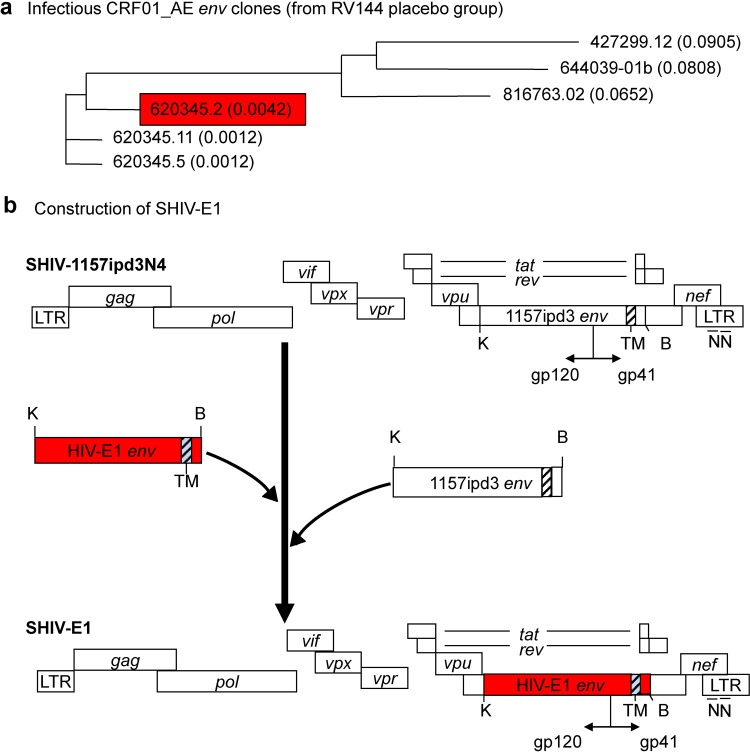
The phase III RV144 human immunodeficiency virus (HIV) vaccine trial conducted in Thailand remains the only study to show efficacy in decreasing the HIV acquisition risk. In Thailand, circulating recombinant forms of HIV clade A/E (CRF01_AE) predominate; in such viruses, env originates from clade E (HIV-E). We constructed a simian-human immunodeficiency virus (SHIV) chimera carrying env isolated from an RV144 placebo recipient in the SHIV-1157ipd3N4 backbone. The latter contains long terminal repeats (LTRs) with duplicated NF-κB sites, thus resembling HIV LTRs. We devised a novel strategy to adapt the parental infectious molecular clone (IMC), R5 SHIV-E1, to rhesus macaques: the simultaneous depletion of B and CD8+ cells followed by the intramuscular inoculation of proviral DNA and repeated administrations of cell-free virus. High-level viremia and CD4+ T-cell depletion ensued. Passage 3 virus unexpectedly caused acute, irreversible CD4+ T-cell loss; the partially adapted SHIV had become dual tropic. Virus and IMCs with exclusive R5 tropism were reisolated from earlier passages, combined, and used to complete adaptation through additional macaques. The final isolate, SHIV-E1p5, remained solely R5 tropic. It had a tier 2 neutralization phenotype, was mucosally transmissible, and was pathogenic. Deep sequencing revealed 99% Env amino acid sequence conservation; X4-only and dual-tropic strains had evolved independently from an early branch of parental SHIV-E1. To conclude, our primate model data reveal that SHIV-E1p5 recapitulates important aspects of HIV transmission and pathobiology in humans.IMPORTANCE Understanding the protective principles that lead to a safe, effective vaccine against HIV in nonhuman primate (NHP) models requires test viruses that allow the evaluation of anti-HIV envelope responses. Reduced HIV acquisition risk in RV144 has been linked to nonneutralizing IgG antibodies with a range of effector activities. Definitive experiments to decipher the mechanisms of the partial protection observed in RV144 require passive-immunization studies in NHPs with a relevant test virus. We have generated such a virus by inserting env from an RV144 placebo recipient into a SHIV backbone with HIV-like LTRs. The final SHIV-E1p5 isolate, grown in rhesus monkey peripheral blood mononuclear cells, was mucosally transmissible and pathogenic. Earlier SHIV-E passages showed a coreceptor switch, again mimicking HIV biology in humans. Thus, our series of SHIV-E strains mirrors HIV transmission and disease progression in humans. SHIV-E1p5 represents a biologically relevant tool to assess prevention strategies.

Keywords: CRF01_AE; HIV; RV144 trial; SHIV-E; adaptation; coreceptor switch; rhesus macaques.


Figures

FIG 1
FIG 1
Construction of SHIV-E1. (A) Several infectious HIV-E1 env clones were identified from newly infected individuals of the RV144 placebo group. env sequence divergence is shown. (B) To generate the initial parental SHIV-E1 provirus, env clone 620345.2 (0.0042) (red box) was inserted into the SHIV-1157ipd3N4 backbone (10). The latter infectious molecular clone had been engineered to contain duplicated NF-κB sites (NN) in the long terminal repeats (LTRs). As such, this SHIV LTR resembles the HIV LTR better than the original SIVmac239 LTR that contains only one NF-κB site. For the sake of simplicity, we use the term “SHIV-E1” for our initial infectious molecular SHIV clone. Of note, in the RV144 CRF01_AE strains used, the env sequences per se originated from HIV clade E strains rather than clade A strains. Thus, the env clone 620345.2 (0.0042) (shown in red boxes in panels A and B) used to generate the SHIV-E1 construct was renamed “HIV-E1 env.” The cloning sites KpnI (K) and BamHI (B) are shown. TM, transmembrane region.
FIG 2
FIG 2
Early passage history of SHIV-E1 in rhesus macaques (RMs). (A) Schema of serial SHIV-E1 passage through different RMs. The first animal, R547, was inoculated intravenously (i.v.) with the cell-free supernatant from 293T cells transfected with the infectious molecular clone SHIV-E1 as well as with SHIV-E1 proviral DNA by the intramuscular (i.m.) route. The second RM, R551, received blood i.v. from animal R547 on day 53. Both animals R547 and R551 were immunodepleted for CD8+ cells and B cells prior to virus exposure (black box). Animal R547 is denoted in red, indicating that the reisolated virus was exclusively R5 tropic. Virus isolated from animal R551 (day 57; day of necropsy) was found to be dual tropic (gradient from red to blue); initial red shading for animal R551 indicates that virus isolated early (day 14) was R5 tropic. Both animals R555 and R561 received dual-tropic virus (blue shading). Solid red arrows indicate the passage direction; olive arrows denote dead-end passages with filtered plasma transfer with dual-tropic virus. Dotted black boxes indicate RMs that were PCR positive for T. cruzi. Circles with P1, P2, P3a, and P3b indicate the passage numbers. (B) Summary of inocula, route of inoculation, and source of the inoculum for each animal.
FIG 3
FIG 3
Replication kinetics of SHIV-E1 in RMs during serial passage. RMs R547 and R551 were immunodepleted by the administration of anti-B cell (rituximab) (green arrows) and anti-CD8+ cell (cM-T807) (black arrows) mAbs on the days indicated. Animal R547 was inoculated with SHIV-E1 IMC DNA i.m. at two sites on day 0 (red asterisk with down arrow) and cell-free virus i.v. on three different days (7 ml each dose) (red down arrows). Blood was transferred to animal R551 on day 53 after inoculation of animal R547. Animal R551 developed signs of AIDS and was euthanized on day 57. Red up arrows indicate the isolation of virus from animals R547 (day 203) and R551 (days 14 and 57); olive green arrows indicate simultaneous, dead-end passages with filtered plasma containing dual-tropic virus from donor R551 to animals R555 and R561 (neither recipient was pretreated with rituximab or cM-T807); circles with P1, P2, P3a, and P3b indicate the passage numbers; red boxes indicate R5-tropic virus; blue boxes indicate dual-tropic virus; dotted black boxes indicate RMs that were PCR positive for T. cruzi; red lines with red circles indicate viral RNA copies per milliliter on a log scale; and axes on the right indicate absolute cell numbers (103).
FIG 4
FIG 4
Coreceptor usage of SHIV-E1p2d57 reisolated from animal R551. (Ai and iv) Infectivity assays for SHIV-E1p2d57 isolated from T. cruzi-infected animal R551 on day 57 using CEMx174-GFP cells. (ii and v) X4-tropic HIV-1NL4-3 was used as a positive control for X4 tropism. (iii and vi) No-virus controls. Top panels represent images acquired in bright field; bottom panels represent the corresponding GFP fluorescence. (B) Virus production measured by a p27 or p24 ELISA of culture supernatants harvested on different days after exposure of U87 or GHOST cells expressing different human coreceptors to cell-free virus preparations. (Left) p27 levels of SHIV-E1p2d57; (middle) p27 levels of the SHIV-E1 parental IMC from transfected 293T cells; (right) p24 levels of HIV-196USSN20, known to have extended coreceptor usage.
FIG 5
FIG 5
Generation of the SHIV-E1p2 infectious molecular clone. (A) Steps involved in the generation of an exclusively R5 SHIV-E1p2 infectious molecular clone. (B) Virus production measured by a p24 ELISA of culture supernatants harvested on day 7 after exposure of U87.CD4.CCR5 or U87.CD4.CXCR4 cells to cell-free supernatants of 293T cells transfected with various NL-LucR.E1p2 IMCs. HIV-1NL4-3 and HIV-11084i were used as X4- and R5-tropic controls, respectively. (C) Infectivity on TZM-bl cells of serially diluted supernatants of 293T cells transfected with various IMCs of SHIV-E1p2. SHIV-KNH1144p4 (SHIV-A) (our unpublished data) was used as a positive control. (D) Ability of PBMC from 7 random naive rhesus macaque donors to support the replication of the SHIV-E1p2c183 IMC. The cell-free filtered supernatant of transfected 293T cells was assayed. Supernatants of IMC-exposed PBMC were collected on the days indicated.
FIG 6
FIG 6
Serial passages P4 and P5 followed by isolation of SHIV-E1p5. (A) Animal R798 was inoculated with cell-free SHIV-E1p1d203 and SHIV-E1p2d14 i.v. and with SHIV-E1p2c183 IMC DNA by the i.m. route. Blood was transferred to animal R974 on day 28 after inoculation of animal R798. SHIV-E1p5 was isolated on day 42 after exposure of animal R974. Red boxes indicate R5-tropic virus. For P1 through P3a and P3b, see the legend to Fig. 2A. (B) Table summarizing the inocula, their sources, and routes of transfer during passages 4 and 5 of SHIV-E1. (C) Virus replication kinetics in animals R798 and R974. Red down arrow with asterisk indicates inoculation of RM R798 with proviral SHIV-E1p2c183 IMC DNA i.m. at two sites on day 0. Red down arrows (without asterisks) indicate inoculation of cell-free virus on three different days. Red up arrow for passage 5 for animal R974 indicates the reisolation of SHIV-E1p5 on day 42 after blood transfer, circles with P4 and P5 indicate the passage numbers, red boxes indicate R5-tropic virus, red lines with red circles indicate viral RNA copies per milliliter on a log scale, and axes on the right indicate absolute cell numbers (103).
FIG 7
FIG 7
SHIV-E1p5 is exclusively R5 tropic. (A) CEMx174-GFP cell infectivity assay with SHIV-E1p5 (i and iv), the X4-tropic positive control HIV-1NL4-3 (ii and v), and R5-tropic SHIV-1157ipd3N4 (iii and vi). (Top) Images acquired in bright field; (bottom) corresponding GFP fluorescence. (B) Infectivity of SHIV-E1p5 in U87.CD4 and GHOST.CD4 cells expressing various coreceptors. HIV-196USSN20 was used as a control.
FIG 8
FIG 8
SHIV-E1p5 replication in vitro and in vivo. (A) Ability of PBMC from six randomly selected naive RM donors to support the replication of SHIV-E1 from passage 1 (SHIV-E1p1d203), passage 2 (SHIV-E1p2d14), and passage 5 (SHIV-E1p5). (B) SHIV-E1p5 replication in PBMC of 10 random naive RM donors. (C) SHIV-E1p5 replication kinetics in a rhesus macaque challenged intrarectally (red down arrow). Plasma vRNA copies per milliliter were assessed by using nucleic acid sequence-based amplification (NASBA) technology. Absolute numbers of blood cells were monitored by FACS analysis. At time zero, the absolute CD4+ T-cell number for the intrarectally challenged animal was 441 cells/mm3, which is within normal limits for adult Indian-origin RMs, according to Autissier et al. (69).
FIG 9
FIG 9
Predicted Env amino acid (AA) changes during the adaptation of SHIV-E1p1d203, SHIV-E1p2d14, the “dead-end” isolate SHIV-E1p2d57, and SHIV-E1p5 by deep sequencing using Illumina technology. The four panels show predicted amino acid changes of the three R5 isolates (black boxes) and dual-tropic SHIV-E1p2d57 (navy blue box) compared to the parental infectious molecular clone, SHIV-E1. The height of each bar represents the percentage of sequence reads containing a given mutation. Sequences with a prevalence of ≥5% are shown. Salmon-colored bars indicate predicted amino acid changes found during adaptation but not associated with a coreceptor switch, and navy blue bars indicate mutations found exclusively in the dual-tropic SHIV-E1p2d57 isolate. CD4i, amino acid mutation G426R, known to be associated with a CD4i epitope and in the region of the chemokine receptor binding site (23, 24). Areas shaded in dark gray indicate Env domains that represent HIV clade E sequences (not drawn to scale), and areas shaded in light gray indicate gp41 regions derived from the SHIV backbone used for cloning, SHIV-1157ipd3N4; these sequences were derived from either HIV clade B or the SIVmac239 Env/Nef overlap (10).
FIG 10
FIG 10
Predicted amino acid changes in the V3 loop of HIV clade E gp120 associated with coreceptor switching. (A) Alignment of V3 loop amino acid sequences of either infectious molecular clones (IMCs) or biological SHIV isolates (designated virus1) with the consensus CRF01_AE sequence (46) and the parental SHIV-E1 IMC. env clones had been inserted into NL-LucR vectors and were shown to be infectious; env clones had been isolated on day 14 or 57 from passage 2. The sequence of SHIV-E1p2d14c183, a clone that was used for subsequent readaptation, is also listed. Sequences starting with EU and GU were derived from data reported previously and are designated reference (Ref) clones (46, 70). A red box indicates V3 loop sequences associated with R5 tropism, and a blue box indicates sequences associated with dual tropism or X4 usage. The tropism was predicted by the PhenoSeq platform (22). (B) SHIV-E1 phylogenetic tree. Env amino acid sequences of the SHIV-E1 parental IMC and SHIV-E1 isolates were aligned and analyzed for evolutionary relatedness in Mega7. The branch lengths reflect the number of substitutions per site. Red boxes indicate R5 viruses, and blue boxes indicate R5X4/X4 viruses.

Similar articles

Cited by

References

    1. Rerks-Ngarm S, Pitisuttithum P, Nitayaphan S, Kaewkungwal J, Chiu J, Paris R, Premsri N, Namwat C, de Souza M, Adams E, Benenson M, Gurunathan S, Tartaglia J, McNeil JG, Francis DP, Stablein D, Birx DL, Chunsuttiwat S, Khamboonruang C, Thongcharoen P, Robb ML, Michael NL, Kunasol P, Kim JH. 2009. Vaccination with ALVAC and AIDSVAX to prevent HIV-1 infection in Thailand. N Engl J Med 361:2209–2220. doi: 10.1056/NEJMoa0908492. - DOI - PubMed
    1. Haynes BF, Gilbert PB, McElrath MJ, Zolla-Pazner S, Tomaras GD, Alam SM, Evans DT, Montefiori DC, Karnasuta C, Sutthent R, Liao HX, DeVico AL, Lewis GK, Williams C, Pinter A, Fong Y, Janes H, DeCamp A, Huang Y, Rao M, Billings E, Karasavvas N, Robb ML, Ngauy V, de Souza MS, Paris R, Ferrari G, Bailer RT, Soderberg KA, Andrews C, Berman PW, Frahm N, De Rosa SC, Alpert MD, Yates NL, Shen X, Koup RA, Pitisuttithum P, Kaewkungwal J, Nitayaphan S, Rerks-Ngarm S, Michael NL, Kim JH. 2012. Immune-correlates analysis of an HIV-1 vaccine efficacy trial. N Engl J Med 366:1275–1286. doi: 10.1056/NEJMoa1113425. - DOI - PMC - PubMed
    1. Bonsignori M, Pollara J, Moody MA, Alpert MD, Chen X, Hwang KK, Gilbert PB, Huang Y, Gurley TC, Kozink DM, Marshall DJ, Whitesides JF, Tsao CY, Kaewkungwal J, Nitayaphan S, Pitisuttithum P, Rerks-Ngarm S, Kim JH, Michael NL, Tomaras GD, Montefiori DC, Lewis GK, DeVico A, Evans DT, Ferrari G, Liao HX, Haynes BF. 2012. Antibody-dependent cellular cytotoxicity-mediating antibodies from an HIV-1 vaccine efficacy trial target multiple epitopes and preferentially use the VH1 gene family. J Virol 86:11521–11532. doi: 10.1128/JVI.01023-12. - DOI - PMC - PubMed
    1. Chung AW, Ghebremichael M, Robinson H, Brown E, Choi I, Lane S, Dugast AS, Schoen MK, Rolland M, Suscovich TJ, Mahan AE, Liao L, Streeck H, Andrews C, Rerks-Ngarm S, Nitayaphan S, de Souza MS, Kaewkungwal J, Pitisuttithum P, Francis D, Michael NL, Kim JH, Bailey-Kellogg C, Ackerman ME, Alter G. 2014. Polyfunctional Fc-effector profiles mediated by IgG subclass selection distinguish RV144 and VAX003 vaccines. Sci Transl Med 6:228ra38. doi: 10.1126/scitranslmed.3007736. - DOI - PubMed
    1. Chung AW, Kumar MP, Arnold KB, Yu WH, Schoen MK, Dunphy LJ, Suscovich TJ, Frahm N, Linde C, Mahan AE, Hoffner M, Streeck H, Ackerman ME, McElrath MJ, Schuitemaker H, Pau MG, Baden LR, Kim JH, Michael NL, Barouch DH, Lauffenburger DA, Alter G. 2015. Dissecting polyclonal vaccine-induced humoral immunity against HIV using systems serology. Cell 163:988–998. doi: 10.1016/j.cell.2015.10.027. - DOI - PMC - PubMed

Publication types

LinkOut - more resources