. 2018 May 9;8(1):7350.
doi: 10.1038/s41598-018-25387-8.

Released Mitochondrial DNA Following Intestinal Ischemia Reperfusion Induces the Inflammatory Response and Gut Barrier Dysfunction

Affiliations

Released Mitochondrial DNA Following Intestinal Ischemia Reperfusion Induces the Inflammatory Response and Gut Barrier Dysfunction

Qiongyuan Hu et al. Sci Rep. .

Erratum in

Abstract

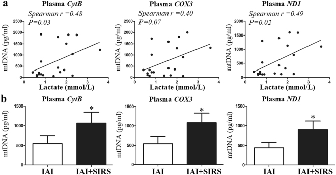
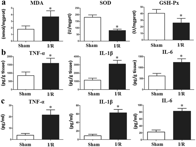
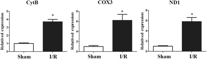
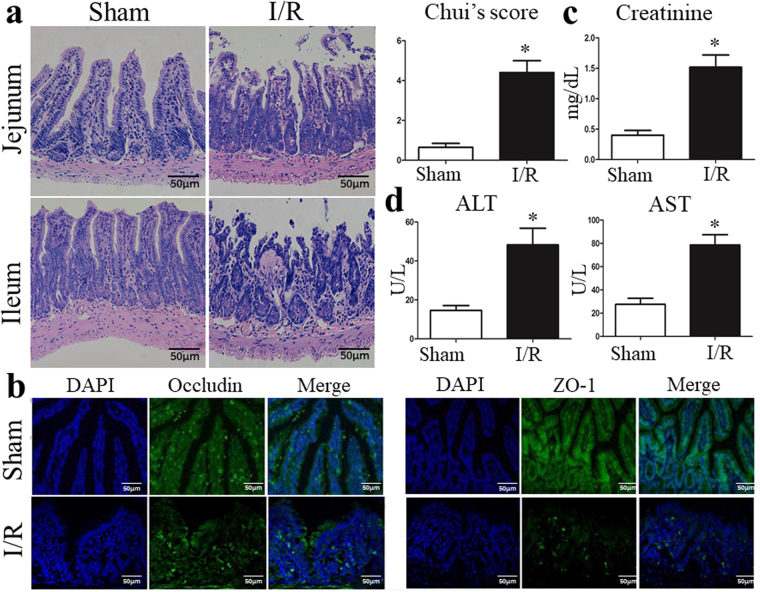
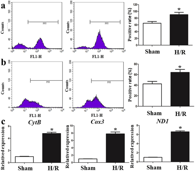
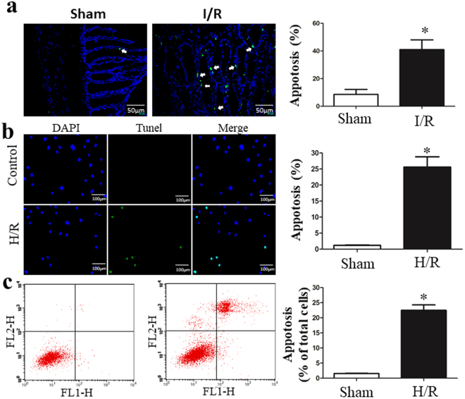
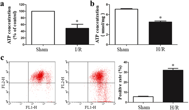
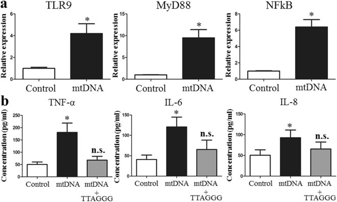
Ischemia-reperfusion (I/R) injury is a challenging clinical problem, especially injuries involving the gastrointestinal tract. Mitochondrial DNA (mtDNA) is released upon cell death and stress, and can induce the inflammatory response. We aimed to investigate the role of mtDNA in the pathogenesis of intestinal I/R. Intestinal I/R model was established with clamping of the superior mesenteric artery, and IEC-6 cells were incubated under hypoxia/reoxygenation (H/R) conditions to simulate I/R injury. Using in vitro models, H/R up-regulated oxidative stress, disrupted mitochondrial activity and the mitochondrial membrane potential, induced apoptosis and elevated the mtDNA levels in the supernatant of intestinal epithelial cells, and the co-culture of mtDNA with human primary dendritic cells significantly elevated TLR9-MyD88 expression and enhanced the production of inflammatory cytokines and chemokines. MtDNA was also released in a mouse model of intestinal I/R and was associated with the increased secretion of inflammatory cytokines and increased gut barrier injury compared with that of the sham group. We concluded that mtDNA contributes to I/R injury and may serve as a biomarker of intestinal I/R. We further suggest that oxidized mtDNA originated from IECs during intestinal I/R exacerbates the acute proinflammatory process by eliciting the production of proinflammatory cytokines and chemokines.


Conflict of interest statement

The authors declare no competing interests.

Figures

Figure 1
Figure 1
The plasma mtDNA level is increased in IAI patients and is correlated with the lactate level. Three selected sequences were used to detect the mtDNA levels using qPCR. (a) Correlations between the baseline mtDNA levels and admission lactate concentration. (b) Plasma mtDNA levels between the IAI group and IAI + SRIS group. mtDNA concentrations are presented as means ± SD. *means P < 0.05.
Figure 2
Figure 2
Total intestinal I/R causes oxidative stress and inflammation in vivo. (a) The levels of SOD and GSH-Px were significantly decreased, and the MDA level was markedly increased in the I/R group compared with that in the sham group. Both the tissue (b) and serum (c) IL-1β, IL-6 and TNF-α levels production were found to be markedly increased after only two hours of intestinal I/R. The Data were expressed as means ± SD. *p < 0.05 vs the I/R group.
Figure 3
Figure 3
Intestinal I/R increases the mtDNA level in the circulation in vivo. Plasma mtDNA levels were significantly elevated in mice undergoing I/R compared with those in the sham group. The data were expressed as means ± SD. *p < 0.05 vs I/R group.
Figure 4
Figure 4
I/R triggers gut injury and intestinal barrier disruption. (a) Representative images of intestinal histology (H&E staining; original magnification, ×200) and histopathological scores (Chiu’s score) of the intestine after intestinal I/R. (b) Localization of occludin and ZO-1, and DAPI staining within intestinal tissue sections as assessed by immunofluorescence at 6 hours after intestinal I/R. The levels of plasma (c) creatinine, (d) ALT and AST were also measured by ELISA at 6 hours after I/R. The data were expressed as means ± SD. *p < 0.05 vs the I/R group.
Figure 5
Figure 5
I/R elevates oxidative stress and mtDNA in vitro. (a) The intracellular ROS levels were increased in H/R-treated IEC-6 cells. (b) H/R treatment obviously elevated mitochondrial ROS in IEC-6 cells. (c) Cells with H/R exposure showed significantly higher mtDNA levels than normoxic controls. The data are expressed as means ± SD. *P < 0.05 vs the H/R group.
Figure 6
Figure 6
I/R induces cell death. (a) Intestinal I/R caused fragmentary or pyknotic-like nuclei, and the chromatin was aggregated in the areas surrounding the nuclei. (b) Representative images and the apoptosis index of in situ TUNEL assay of IEC-6 cells. TUNEL-positive is green and DAPI is blue. (c) AnnexinV/PI analysis with flow cytometry confirmed that H/R-induced cell death. Data are expressed as mean ± SD. *P < 0.05 vs H/R group.
Figure 7
Figure 7
I/R caused mitochondrial dysfunction. The ATP concentration was quantified using a commercially available kit. (a) The ATP concentrations were significantly decreased in the I/R group in vivo. (b) H/R decreased the ATP levels compared with those in the control group. (c) The H/R-treated IEC-6 cells showed a decrease in the mitochondrial membrane potential (ΔΨm). The data are expressed as means ± SD. *P < 0.05 vs the sham group or H/R group.
Figure 8
Figure 8
mtDNA induces pDC cell lines to up-regulate TLR9 expression and induced the inflammatory response. (a) The expression levels of TLR9, MyD88, and NF-κB was significantly increased with mtDNA co-culture compared with those in the control. (b) Freshly isolated primary human pDCs were treated with oxidatively modified mtDNA in the absence or presence of a TLR9 antagonist (TTAGGG). The data are expressed as means ± SD. *P < 0.05 vs the sham group or H/R group.

References

    1. Li Y, et al. 6-Gingerol protects intestinal barrier from ischemia/reperfusion-induced damage via inhibition of p38 MAPK to NF-kappaB signalling. Pharmacological research. 2017;119:137–148. doi: 10.1016/j.phrs.2017.01.026. - DOI - PubMed
    1. Meszaros, A. T. et al. Inhalation of methane preserves the epithelial barrier during ischemia and reperfusion in the rat small intestine. Surgery, 10.1016/j.surg.2016.12.040 (2017). - PubMed
    1. Tassopoulos A, Chalkias A, Papalois A, Iacovidou N, Xanthos T. The effect of antioxidant supplementation on bacterial translocation after intestinal ischemia and reperfusion. Redox Report. 2017;22:1–9. doi: 10.1080/13510002.2016.1229893. - DOI - PMC - PubMed
    1. Mittal R, Coopersmith CM. Special issue: Sepsis Redefining the gut as the motor of critical illness. Trends in Molecular Medicine. 2014;20:214–223. doi: 10.1016/j.molmed.2013.08.004. - DOI - PMC - PubMed
    1. de Jong, P. R., Gonzalez-Navajas, J. M. & Jansen, N. J. G. The digestive tract as the origin of systemic inflammation. Critical Care20, 10.1186/s13054-016-1458-3 (2016). - PMC - PubMed

Publication types

MeSH terms

Substances

LinkOut - more resources