. 2018 May 9;8(1):7381.
doi: 10.1038/s41598-018-25765-2.

Substrate specificity of human MCPIP1 endoribonuclease

Affiliations

Substrate specificity of human MCPIP1 endoribonuclease

Mateusz Wilamowski et al. Sci Rep. .

Abstract

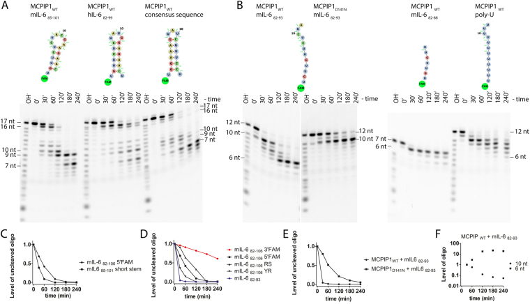
MCPIP1, also known as Regnase-1, is a ribonuclease crucial for regulation of stability of transcripts related to inflammatory processes. Here, we report that MCPIP1 acts as an endonuclease by degrading several stem-loop RNA structures and single-stranded RNAs. Our studies revealed cleavage sites present in the stem-loops derived from the 3' untranslated region of the interleukin-6 transcript. Furthermore, MCPIP1 induced endonuclease cleavage at the loop motif of stem-loop structures. Additionally, we observed that MCPIP1 could cleave single-stranded RNA fragments. However, RNA substrates shorter than 6 nucleotides were not further affected by MCPIP1 nucleolytic activity. In this study, we also determined the dissociation constants of full-length MCPIP1D141N and its ribonuclease domain PIN D141N with twelve oligonucleotides substrates. The equilibrium binding constants (Kd) for MCPIP1D141N and the RNA targets were approximately 10 nM. Interestingly, we observed that the presence of a zinc finger in the PIN domain increases the affinity of this protein fragment to 25-nucleotide-long stem-loop RNA but not to shorter ones. Furthermore, size exclusion chromatography of the MCPIP1 and PIN proteins suggested that MCPIP1 undergoes homooligomerization during interaction with RNA substrates. Our results provide insight into the mechanism of MCPIP1 substrate recognition and its affinity towards various oligonucleotides.


Conflict of interest statement

The authors declare no competing interests.

Figures

Figure 1
Figure 1
RNA fragments obtained upon MCPIP1-catalyzed cleavage. (A) Sequences and structures of the 25-nt-long RNA stem-loops derived from the mIL-682–106 3′UTR fragment are depicted. (B) Degradation assay of the modified mIL-682–106 stem-loop (RS – reverse stem alteration). (C) Degradation assay of the modified mIL-682–106 stem-loop (YR – pyrimidine and purine alterations). (D) Degradation assay of the mIL-682–106 stem-loop labeled at the 3′ end. The oligonucleotides were labeled at the 5′ or 3′ end with FAM dye. The reaction products were resolved on 20% denaturing PAGE and visualized with a fluorescence imaging system. Concentrations of the labeled oligonucleotides and MCPIP1 protein were 7.5 μM and 2 μM, respectively. The D141N mutation of a conserved aspartate of the PIN domain catalytic center of the MCPIP1 decreased its ribonucleolytic activity. (AD) Were made by the separation of gels that are shown at Supplementary Fig. S2A,D. Major sites of MCPIP1-induced cleavage are indicated by the oligonucleotide fragment length. (E) Densitometric analysis of the kinetics of RNA degradation presented as the level of the remaining uncleaved oligonucleotides from the RNase assay. The graph shows the level of the stem-loop sequence: mIL-682–106 RS during degradation. (F) Densitometric analysis of the MCPIP1WT induced degradation of the mIL-682–106 5′FAM oligonucleotide. Analysis was carried out separately for each of degradation products of the mIL-682–106 5′FAM. Oligonucleotide levels were normalized at the time 30 min.
Figure 2
Figure 2
RNA fragments obtained upon cleavage catalyzed by MCPIP1. Major sites of MCPIP1-induced cleavage are indicated by the oligonucleotide fragment length. (A) Sequences and structures of the short stem-loops are depicted. These sequences form 17-18-nt-long stem-loops. (B) Degradation of the single-stranded RNAs. Twelve and 7-nt-long single-stranded RNA sequences were part of the mIL-682–106 stem-loop. Poly-U homopolymer RNA contains 12 uracil residues. The oligonucleotides were labeled at the 5′ end with FAM dye. The reaction products were resolved on 20% denaturing PAGE and visualized with a fluorescence imaging system. Concentrations of the labeled oligonucleotides and MCPIP1 protein were 7.5 μM and 2 μM, respectively. The D141N mutation of a conserved aspartate of the PIN domain catalytic center of the MCPIP1 decreased its ribonucleolytic activity. (A,B) Were made by the separation of gels that are shown at Supplementary Fig. S2B–D. (C) Densitometric analysis of the level of the remaining uncleaved oligonucleotides from the RNase assay at different time points. The panel shows a comparison of the kinetics of degradation of the mIL-682–106 and a shorter fragment of this stem-loop, which is mIL-685–101. (D) The panel shows the level of degradation of the 25-nt-long stem-loop mIL-682–106 in comparison with the 12 nt ssRNA sequence of the mIL-682–93. (E) The graph compares the degradation of the ssRNA mIL682–93 with MCPIP1WT or MCPIP1D141N, which possesses attenuated RNase activity. (F) Densitometric analysis of the MCPIP1WT induced degradation of the mIL-682–93 oligonucleotide. The analysis was carried out for 6 nt and 10 nt long mIL-682–93 degradation products respectively, oligonucleotide levels were normalized at the time 30 min.
Figure 3
Figure 3
DNA fragments obtained upon cleavage catalyzed by MCPIP1. (A) The DNA sequences are based on the mIL-682–106 sequence; one oligonucleotide is ssDNA, and the second is dsDNA. The oligonucleotides were labeled at the 5′ end with FAM dye. The reaction products were resolved on 20% denaturing PAGE and visualized with a fluorescence imaging system. Concentrations of the labeled oligonucleotides and MCPIP1 protein were 7.5 μM and 2 μM, respectively. The D141N mutation of a conserved aspartate of the PIN domain catalytic center of the MCPIP1 decreased its ribonucleolytic activity. (A) Was made by the separation of gels that are shown at Supplementary Fig. S2C,D. (B) Densitometric analysis of the level of the remaining uncleaved ssDNA and dsDNA oligonucleotides from the MCPIP1 nuclease activity assay at different time points.
Figure 4
Figure 4
(A) Domain characterization of MCPIP1: UBA43–89 (Ubiquitin-associated domain); PRR100–126 and 458–536 (Proline-rich region); PIN133–270 (PilT N-terminus nuclease domain); ZF305–325 (zinc-finger motif); disordered region326–457; CTD545–598 (C-terminal conserved domain). Depicted fragments of MCPIP1, PIN-ZF and PIN that were used in presented studies. (B) Affinity of the MCPIP1 interaction with oligonucleotides forming RNA stem-loop structures: mIL-682–106 5′FAM and single-stranded RNA oligonucleotides represented by mIL-682–93. The analyzed proteins were the catalytic mutated forms: MCPIP1D141N and its PIN-ZFD141N and PIND141N fragments. The ribonuclease PIND141N domain was studied without or with the zinc finger motif at the C-terminal region. Graphs illustrate the interaction of selected proteins (MCPIP1D141N, PIN-ZFD141N, and PIND141N) with oligonucleotides. Functions were fitted to the fluorescence intensity data points using the sequential binding model N + P + P NP + P NPP (P – protein N – oligonucleotide). The depicted errors bars are standard deviations, n = 3. (C) Controls of the affinity determination assay. MCPIP1D141N in a presence of the free FAM label and unlabeled hIL-681–98 RNA oligonucleotide.
Figure 5
Figure 5
Homooligomerization of the MCPIP1 protein. (A) Size exclusion chromatography results of MCPIP1. Chromatography was performed in a buffer comprised of 25 mM Tris, pH 7.9, 300 mM NaCl, 10% (w/v) glycerol, 1 mM DTT, and 0.5 mM EDTA. Additionally, for results shown as a dotted line, the buffer was enriched in 1.6 M urea. (B) A multiple Gaussian peak fit was performed to model the obtained elution profile of MCPIP1WT. Fitted peaks illustrated tetrameric, dimeric and monomeric fractions of the MCPIP1WT. (C) Calibration curve of the gel filtration column. Green points indicate the apparent molecular weight of the investigated proteins calculated using the calibration curve. The molecular weights of these proteins are as follows: MCPIP1: 65.7 kDa; PIN-ZF: 24.7 kDa; PIN: 21.1 kDa. (D) Percentages of the MCPIP1WT tetrameric, dimeric, and monomeric fractions were calculated based on the area of the size exclusion chromatography peaks. (E) Native PAGE results of MCPIP1WT sample in buffers containing 50 mM Tris-HCl, pH 8.3, 150 mM NaCl, 10% (w/v) glycerol, 2.5 mM MgCl2, 1 mM DTT, 0.5 mM EDTA and 0.05 mM ZnCl2. Additional buffer condition changes were an increased concentration of NaCl to 500 mM and addition of urea to 1600 mM.
Figure 6
Figure 6
(A) Identification of MCPIP1-triggered cleavage sites in the mIL-682–106 stem-loop RNA structure. Nt sequences and structures created during MCPIP1-induced cleavage are illustrated. Mapping of the cleavage sites based on the RNase assay results, intermediates and the most significant subsequent degradation products are presented. (B) Visualization of the stoichiometry of the MCPIP1 interaction with stem-loops. Schematic cartoon representation of the ternary complex model. The size exclusion chromatography results showed that PIN and PIN-ZF were monomeric and suggest that full-length MCPIP1 most frequently occurs as a dimer in native condition. Stoichiometry of the MCPIP1 - RNA interaction was based on the size exclusion chromatography results and the results from affinity determination assays where the sequential binding model were used. Thus, for full-length MCPIP1, we proposed a sequential binding model: oligo + MCPIP1Dimer + MCPIP1dimer oligo-MCPIP1dimer + MCPIP1dimer oligo-MCPIP1tetramer. The presented dissociations constants of the complexes were estimated based on the affinity determination assays shown in Table 2.

References

    1. Mizgalska D, et al. Interleukin-1-inducible MCPIP protein has structural and functional properties of RNase and participates in degradation of IL-1β mRNA. FEBS J. 2009;276:7386–7399. doi: 10.1111/j.1742-4658.2009.07452.x. - DOI - PubMed
    1. Matsushita K, et al. Zc3h12a is an RNase essential for controlling immune responses by regulating mRNA decay. Nature. 2009;458:1185–1190. doi: 10.1038/nature07924. - DOI - PubMed
    1. Iwasaki H, et al. The IκB kinase complex regulates the stability of cytokine-encoding mRNA induced by TLR–IL-1R by controlling degradation of regnase-1. Nat. Immunol. 2011;12:1167–1175. doi: 10.1038/ni.2137. - DOI - PubMed
    1. Li M, et al. MCPIP1 down-regulates IL-2 expression through an ARE-independent pathway. PLoS One. 2012;7:e49841. doi: 10.1371/journal.pone.0049841. - DOI - PMC - PubMed
    1. Mino T, et al. Regnase-1 and roquin regulate a common element in inflammatory mRNAs by spatiotemporally distinct mechanisms. Cell. 2015;161:1058–1073. doi: 10.1016/j.cell.2015.04.029. - DOI - PubMed

Publication types