. 2018 May 9;8(1):7355.
doi: 10.1038/s41598-018-25688-y.

Combination of anti-vascular agent - DMXAA and HIF-1α inhibitor - digoxin inhibits the growth of melanoma tumors

Affiliations

Combination of anti-vascular agent - DMXAA and HIF-1α inhibitor - digoxin inhibits the growth of melanoma tumors

Ryszard Smolarczyk et al. Sci Rep. .

Erratum in

Abstract

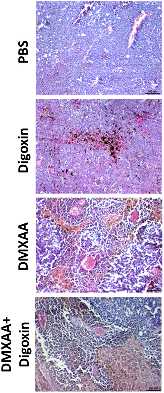
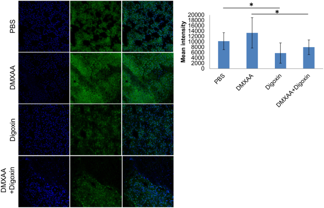
Vascular disrupting agents as DMXAA inhibit tumor growth only for a short period of time followed by rapid tumor regrowth. Among others, hypoxia and presence of transcription factor HIF-1α are responsible for tumors regrowth. The aim of our study was to investigate the inhibition of murine melanoma growth by combining two agents: anti-vascular - DMXAA and the HIF-1α inhibitor - digoxin and explaining the mechanism of action of this combination. After DMXAA treatment tumor size was reduced only for a limited time. After 7 days regrowth of tumors was observed and number of vessels was increased especially in tumor's peripheral areas. DMXAA also induced an influx of immune cells: macrophages, CD8+ cytotoxic lymphocytes, NK cells, CD4+ lymphocytes. Administration of digoxin alone inhibited the growth of tumors. Administration of both agents in the proper sequence significantly inhibited the regrowth of tumors better than either agents alone. Combination therapy reduced number of newly formed vessels. In tumors of mice treated with combination therapy, the number of macrophages M1, CD8+ cytotoxic lymphocytes, NK cells and to a lesser extent CD4+ cells was increased. The combination of anti-vascular agents with HIF-1α inhibitors appears to be an effective therapeutic option.


Conflict of interest statement

The authors declare no competing interests.

Figures

Figure 1
Figure 1
Treatment of mice bearing B16-F10 tumors with DMXAA and digoxin. DMXAA was administered once (25 mg/kg) and digoxin 7 times (2 mg/kg). Inhibition of tumor growth was statistically significant (*p < 0,05) after administration of combined therapy.
Figure 2
Figure 2
Hematoxylin and eosin staining after administration of DMXAA and digoxin. After administration of DMXAA and combined therapy destroyed blood vessels and infiltration of immune cells in the tumors were observed. Digoxin was shown to reduce the area of necrosis in tumors. Lens magnification 10×.
Figure 3
Figure 3
The area occupied by blood vessels in tumors after treatment with DMXAA and digoxin. DMXAA significantly decreases the area of blood vessels in the tumor (*p < 0,05). After digoxin administration it is only slightly reduced. 7 days after DMXAA administration the vessels area increases while in the group treated with the combined therapy it is significantly smaller.
Figure 4
Figure 4
HIF-1α identification in tumors. The amount of HIF-1α factor decreased in tumors of mice that received digoxin and combined therapy compared to mice that received PBS and DMXAA (p < 0.05). In the group of mice that received DMXAA, the amount of HIF-1α increased (no statistical significance).
Figure 5
Figure 5
Identification of macrophages in murine melanoma after administration of DMXAA and digoxin. Cytotoxic (M1) macrophages F4/80+/CD206 infiltration was observed after administration of DMXAA and digoxin. Digoxin alone does not alter the level of macrophages F4/80+/CD206+ (M2) and F4/80+/CD206 (M1).
Figure 6
Figure 6
Presence of immune cells in murine melanoma after administration of DMXAA and digoxin. DMXAA alone and the combination of DMXAA with digoxin increase the levels of CD8+ lymphocytes, NK cells and to a lesser extent CD4+ lymphocytes (A). Digoxin alone does not change CD8+, CD4+, NK cells contribution comparing to control (PBS). Representative plots of cytometric analysis (B).
Figure 7
Figure 7
Scheme of action of the drugs. DMXAA administration to mice bearing tumors leads to the destruction of existing tumor blood vessels. Destruction of the vessels results in the formation of hypoxia areas where HIF-1α transcription factor expression is increased. Activation of HIF-1α triggers several mechanisms that promote tumor growth. Inhibition of HIF-1α after digoxin administration supresses tumor regrowth.

References

    1. Mita MM, Sargsyan L, Mita AC, Spear M. Vascular-disrupting agents in oncology. Expert Opin. Investig. Drugs. 2013;22:317–328. doi: 10.1517/13543784.2013.759557. - DOI - PubMed
    1. Kim S, Peshkin L, Mitchison TJ. Vascular disrupting agent drug classes differ in effects on the cytoskeleton. PLoS One. 2012;7:e40177. doi: 10.1371/journal.pone.0040177. - DOI - PMC - PubMed
    1. Su M, et al. The anti-angiogenic effect and novel mechanisms of action of combretastatin A-4. Sci. Rep. 2016;6:28139. doi: 10.1038/srep28139. - DOI - PMC - PubMed
    1. Surpris G, Poltorak A. The expanding regulatory network of STING-mediated signaling. Curr. Opin. Microbiol. 2016;32:144–150. doi: 10.1016/j.mib.2016.05.014. - DOI - PMC - PubMed
    1. Poltorak A, Kurmyshkina O, Volkova T. Stimulator of interferon genes (STING): a “new chapter” in virus-associated cancer research. Lessons from wild-derived mouse models of innate immunity. Cytokine Growth Factor Rev. 2016;29:83–91. doi: 10.1016/j.cytogfr.2016.02.009. - DOI - PubMed

Publication types

MeSH terms