. 2018 Aug;23(8):1699-1710.
doi: 10.1038/s41380-018-0053-1. Epub 2018 May 9.

Synaptic phospholipids as a new target for cortical hyperexcitability and E/I balance in psychiatric disorders

Affiliations

Synaptic phospholipids as a new target for cortical hyperexcitability and E/I balance in psychiatric disorders

Carine Thalman et al. Mol Psychiatry. 2018 Aug.

Erratum in

Abstract

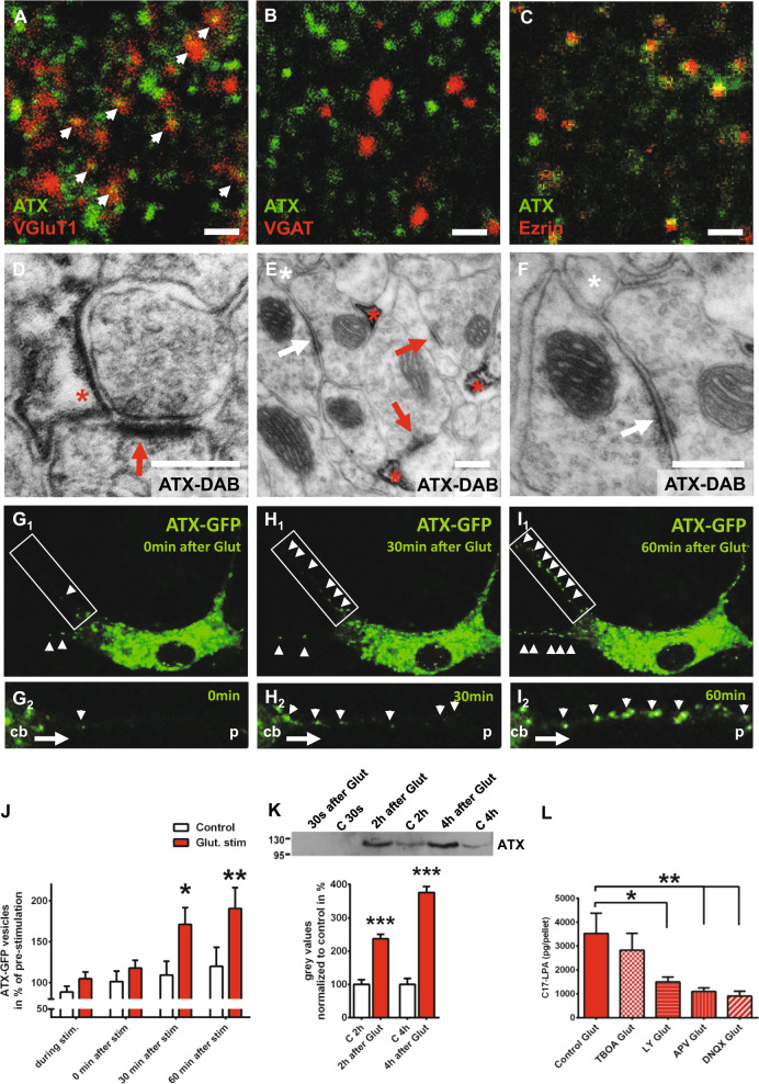
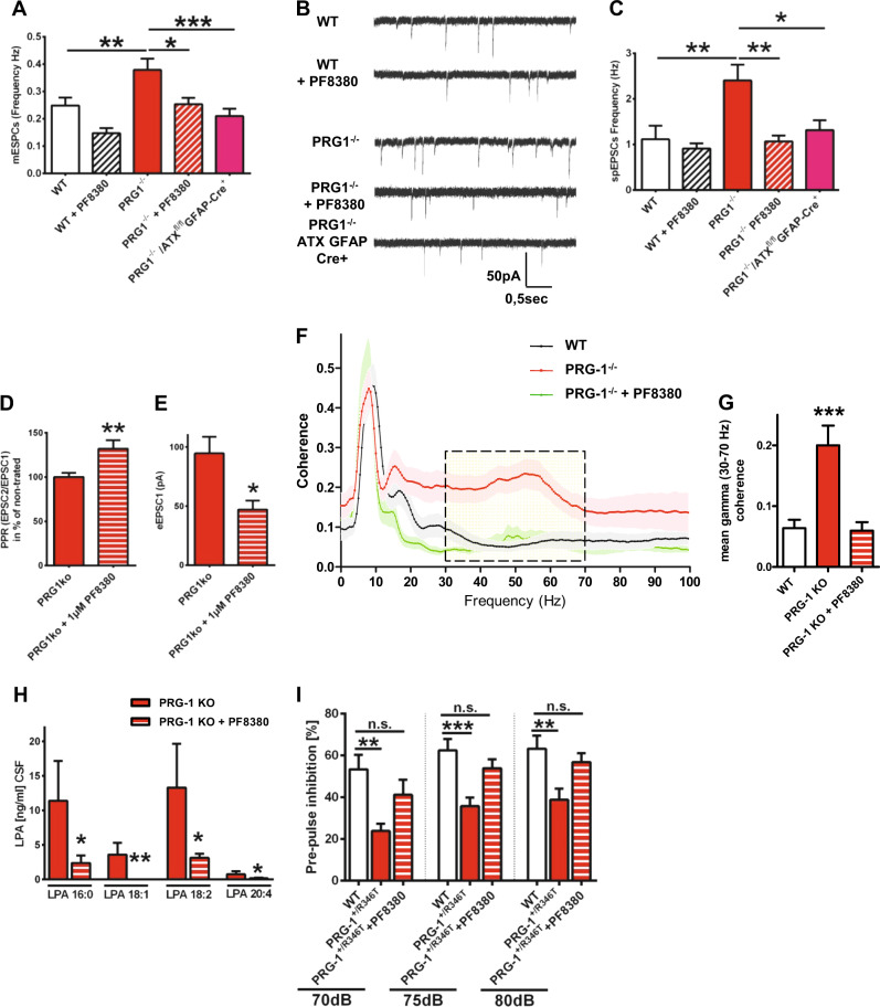
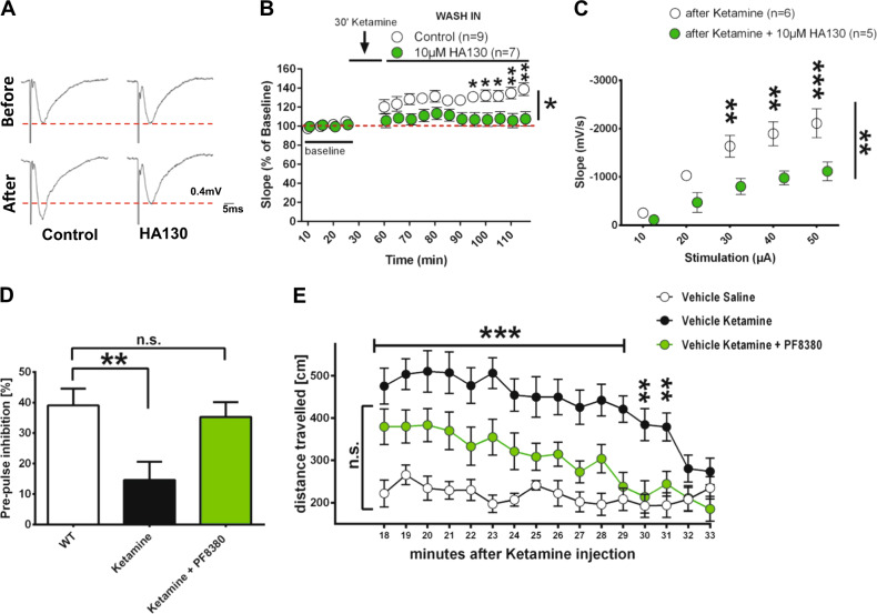
Lysophosphatidic acid (LPA) is a synaptic phospholipid, which regulates cortical excitation/inhibition (E/I) balance and controls sensory information processing in mice and man. Altered synaptic LPA signaling was shown to be associated with psychiatric disorders. Here, we show that the LPA-synthesizing enzyme autotaxin (ATX) is expressed in the astrocytic compartment of excitatory synapses and modulates glutamatergic transmission. In astrocytes, ATX is sorted toward fine astrocytic processes and transported to excitatory but not inhibitory synapses. This ATX sorting, as well as the enzymatic activity of astrocyte-derived ATX are dynamically regulated by neuronal activity via astrocytic glutamate receptors. Pharmacological and genetic ATX inhibition both rescued schizophrenia-related hyperexcitability syndromes caused by altered bioactive lipid signaling in two genetic mouse models for psychiatric disorders. Interestingly, ATX inhibition did not affect naive animals. However, as our data suggested that pharmacological ATX inhibition is a general method to reverse cortical excitability, we applied ATX inhibition in a ketamine model of schizophrenia and rescued thereby the electrophysiological and behavioral schizophrenia-like phenotype. Our data show that astrocytic ATX is a novel modulator of glutamatergic transmission and that targeting ATX might be a versatile strategy for a novel drug therapy to treat cortical hyperexcitability in psychiatric disorders.


Conflict of interest statement

The authors declare that they have no conflict of interest.

Figures

Fig. 1
Fig. 1
ATX is expressed by perisynaptic astrocytic processes (PAPs) at excitatory synapses and is regulated by glutamate. a, b ATX shows abundant colocalization with VGluT1, a marker of excitatory synapses, but not with VGAT, a marker of inhibitory synapses. c ATX is expressed at PAPs as shown by colocalization with Ezrin, a specific marker for PAPs. Scale bars a–c: 1 µm. d Electron microscopic image showing ATX expression (displayed by DAB-precipitation [red asterisk] at the astrocytic membrane close to the synapse) on PAP membranes surrounding glutamatergic synapses. Dense membrane in the postsynaptic compartment (red arrow) represents the postsynaptic density (PSD) of glutamatergic synapses (see also Fig. S1B). e Immune electron microscopic analysis revealed no ATX expression in astrocytic processes (white asterisk) at inhibitory synapses (white arrow). In contrast, ATX expression (shown by DAB precipitation) was prominent in astrocytic processes (red asterisk) at excitatory synapses (red arrow, see also Fig. S1C,D). f Higher magnification showing the inhibitory synapse (white arrow) and the adjacent ATX-negative astrocytic process (white asterisk). Scale bars d–f: 200 nm. G1-I1 Live imaging of astrocytes transfected with an ATX-GFP-expressing construct shows a clear increase of ATX-GFP-positive vesicles transported along their processes towards the periphery after glutamate stimulation (500 µM for 15 min). G2-I2 Live-imaging pictures at higher magnification show transport of ATX-GFP-positive vesicles (white arrow heads) along an astrocytic process from the cell body (cb) toward the periphery (p). In contrast, ATX-GFP-positive vesicles were rarely seen in processes of control, non-stimulated astrocytes (Fig. S1E). j Quantitative analysis of ATX-GFP vesicles in astrocytic processes, during glutamate stimulation and at 0, 30 and 60 min after glutamate stimulation (n = 13 control astrocytes and 14 glutamate-stimulated astrocytes; two-way RM ANOVA with Bonferroni post hoc). k ATX secretion in the astrocytic culture supernatant was significantly increased upon glutamate stimulation (500 µM Glut stimulation for 15 min) when compared with supernatant from control (c), non-stimulated astrocytic cultures as shown by western blot (n = 5 for 2 h values and n = 4 for 4 h values, two-tailed t-test). l C17-LPA synthesis in the astrocytic compartment was significantly decreased upon application of specific glutamate receptor inhibitors (50 µM LY367385 for mGluR1a, 50 µM APV for NMDA-Rs and 10 µM DNQX for AMPA-Rs), whereas 40 µM DL-TBOA, an inhibitor of astrocytic glutamate transporters, did not affect astrocytic C17-LPA levels as shown by mass spectrometry analyses (n = 6 experiments per group, one-way RM ANOVA with Bonferroni post hoc). Bars represent mean ± SEM. *p < 0.05, **p < 0.01, ***p < 0.001
Fig. 2
Fig. 2
ATX inhibition rescues neuronal hyperexcitability and PPI deficits in PRG-1-deficient animals. a Miniature excitatory postsynaptic currents (mEPSCs) in CA1 pyramidal neurons of wild-type (WT) slices were not affected by ATX inhibition with 1 µM PF8380. However, in PRG-1-/- slices, which display higher mEPSC frequency, ATX inhibition significantly diminished mEPSC frequency to WT levels. In line, astrocyte-specific ATX deletion significantly decreased mEPSC frequency in PRG-1-/- animals to WT levels. Amplitudes (see Fig. S2C) were not affected supporting a presynaptic effect (n = 13 WT neurons, 8 PF8380-treated WT neurons, 10 PRG-1-/- neurons, 10 PF8380-treated PRG-1-/- neurons and 10 PRG-1-/-/ATXfl/fl:GFAP-Cre+ neurons; one-way ANOVA with Bonferroni post hoc). b Original traces showing spontaneous excitatory postsynaptic currents (spEPSCs) of neurons under different conditions. c Although ATX inhibition via PF8380 application did not alter spontaneous excitatory (spEPSCs) frequency in WT slices, higher spontaneous frequency observed in PRG-1-/- neurons was significantly decreased to WT levels. Similarly, genetic deletion of ATX in astrocytes decreased spontaneous excitatory frequency in PRG-1-/- neurons to WT levels, whereas amplitudes were not affected (see Fig. S2D) (n = 11 WT neurons, 12 PF8380-treated WT neurons, 19 PRG-1-/- neurons, 17 PF8380-treated PRG-1-/- neurons and 18 PRG-1-/-/ATXfl/fl:GFAP-Cre+ neurons; Kruskal–Wallis test with Dunn’s multiple comparisons post hoc test). d Paired-pulse ratio (PPR) in PRG-1-/- animals was significantly increased after ATX inhibition. e ATX inhibition by PF8380 decreased the first eEPSC(1) in PRG-1-/- animals. f Coherence analysis of simultaneous recordings of field potentials in layer II/III of the medial entorhinal cortex (MEC) and the hippocampal CA1 in freely moving mice revealed significantly higher coherence in the gamma range (30–70 Hz, dashed box) in PRG-1-/- mice. Interestingly, LPA reduction by application of the ATX inhibitor PF8380 reduced this coherence in PRG-1-/- mice to WT level (see also Fig. S3A, D-F). g Quantitative analysis revealed significantly increased mean gamma coherence in PRG-1-/- mice, which was reduced to WT levels upon PF8380-treatment (n = 15 WT mice, 12 untreated PRG1-/- and 7 PF8380-treated PRG1-/- mice; one-way ANOVA with Bonferroni post hoc; see also Fig S3B). h In order to assess the efficiency of ATX inhibition by PF8380, LPA levels were measured in the CSF after PF8380 application (30 mg/kg body weight). Here, we found a clear decrease in the main LPA species (LPA 16:0, 18:1, 18:2 and 20:4; n = 6 for all groups; one tailed, Mann–Whitney test; see also Fig. S2C). i Pre-pulse inhibition (PPI) at all tested loudness levels was significantly decreased in a mouse line expressing a monoallelic human PRG-1 single-nucleotide polymorphism (SNP). However, PF8380 application rescued decreased PPI to WT levels (n = 9 WT mice, 15 untreated PRG1+/R346T and 15 PF8380-treated PRG1+/R346T mice; one-way ANOVA with Bonferroni post hoc; see also Fig. S3G-I). Bars represent mean ± SEM. *p < 0.05, **p < 0.01, ***p < 0.001
Fig. 3
Fig. 3
ATX inhibition effectively reduced cortical hyperexcitability, PPI and hyperlocomotion in an animal model for schizophrenia. a Representative evoked field potentials of control and 10 µM HA130-treated slices before and after ketamine stimulation. Note the potentiation after ketamine application and the rescued field potentials after HA130 application on ketamine-treated slices. Dotted red lines depict control levels. b After an initial depression, ketamine application leads to a significant neuronal potentiation as shown by evoked field potential recordings (1 stimulation every 30 s; for amplitudes see Fig. S4B). However, ATX inhibition by HA130 significantly reduced ketamine potentiation back to baseline levels (n = 8 Ketamine and 7 Ketamine + HA130 (10 µM)-treated slices; two-way RM ANOVA and Bonferroni multiple comparison post hoc test). c Input/output (I/O) assessment of slopes using increasing stimulus intensities shows that HA130 application significantly decreased I/O-relationship (n = 6 Ketamine and 5 ketamine + HA130 (10 µM)-treated slices, two-way RM ANOVA and Bonferroni multiple comparison post hoc test; see also Fig. S4C). d Pre-pulse inhibition (PPI) was significantly reduced in ketamine-treated animals but restored to WT levels by ATX inhibition via in vivo application of PF8380. Startle amplitudes were not altered by ketamine application or by ATX inhibition (n = 13 control animals, 13 ketamine-treated animals and 16 ketamine + PF8380-treated animals; one-way ANOVA with Bonferroni’s multiple comparisons test; see also Fig. S4D) e Ketamine induced significant hyperlocomotion in an open field (OF) setting, whereas in vivo inhibition of ATX via PF8380 administration reduced ketamine-induced hyperlocomotion to WT levels (n = 14 control animals, 14 ketamine-treated animals and 15 ketamine + PF8380-treated animals; two-way RM ANOVA and Sidak’s multiple comparison post hoc test). Bars represent mean ± SEM. *p < 0.05, **p < 0.01, ***p < 0.001

References

    1. Turrigiano G. Too many cooks? Intrinsic and synaptic homeostatic mechanisms in cortical circuit refinement. Annu Rev Neurosci. 2011;34:89–103. doi: 10.1146/annurev-neuro-060909-153238. - DOI - PubMed
    1. Harrison PJ, Weinberger DR. Schizophrenia genes, gene expression, and neuropathology: on the matter of their convergence. Mol Psychiatry. 2005;10:40–68. doi: 10.1038/sj.mp.4001558. - DOI - PubMed
    1. Belforte JE, Zsiros V, Sklar ER, Jiang Z, Yu G, Li Y, et al. Postnatal NMDA receptor ablation in corticolimbic interneurons confers schizophrenia-like phenotypes. Nat Neurosci. 2010;13:76–83. doi: 10.1038/nn.2447. - DOI - PMC - PubMed
    1. Trimbuch T, Beed P, Vogt J, Schuchmann S, Maier N, Kintscher M, et al. Synaptic PRG-1 modulates excitatory transmission via lipid phosphate-mediated signaling. Cell. 2009;138:1222–35. doi: 10.1016/j.cell.2009.06.050. - DOI - PMC - PubMed
    1. Vogt J, Yang JW, Mobascher A, Cheng J, Li Y, Liu X, et al. Molecular cause and functional impact of altered synaptic lipid signaling due to a prg-1 gene SNP. EMBO Mol Med. 2016;8:25–38. doi: 10.15252/emmm.201505677. - DOI - PMC - PubMed

Publication types

MeSH terms