. 2018 Mar 18:2018:1975167.
doi: 10.1155/2018/1975167. eCollection 2018.

Apoptosis and Mobilization of Lymphocytes to Cardiac Tissue Is Associated with Myocardial Infarction in a Reperfused Porcine Model and Infarct Size in Post-PCI Patients

Affiliations

Apoptosis and Mobilization of Lymphocytes to Cardiac Tissue Is Associated with Myocardial Infarction in a Reperfused Porcine Model and Infarct Size in Post-PCI Patients

Maria J Forteza et al. Oxid Med Cell Longev. .

Abstract

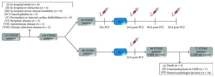
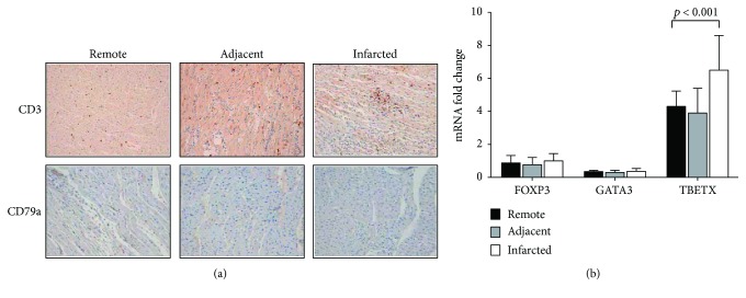
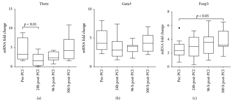
ST-segment elevation myocardial infarction (STEMI) is the most severe outcome of coronary artery disease. Despite rapid reperfusion of the artery, acute irrigation of the cardiac tissue is associated with increased inflammation. While innate immune response in STEMI is well described, an in-depth characterization of adaptive immune cell dynamics and their potential role remains elusive. We performed a translational study using a controlled porcine reperfusion model of STEMI and the analysis of lymphocyte subsets in 116 STEMI patients undergoing percutaneous coronary intervention (PCI). In the animal model, a sharp drop in circulating T lymphocytes occurred within the first hours after reperfusion. Notably, increased apoptosis of circulating lymphocytes and infiltration of proinflammatory Th1 lymphocytes in the heart were observed 48 h after reperfusion. Similarly, in STEMI patients, a sharp drop in circulating T lymphocyte subsets occurred within the first 24 h post-PCI. A cardiac magnetic resonance (CMR) evaluation of these patients revealed an inverse association between 24 h circulating T lymphocyte numbers and infarction size at 1-week and 6-month post-PCI. Our translational approach revealed striking changes in the circulating and tissue-infiltrating T lymphocyte repertoire in response to ischemia-reperfusion. These findings may help in developing new diagnostic and therapeutic approaches for coronary diseases.


Figures

Figure 1
Figure 1
Flowchart of the study group. This flowchart illustrates the STEMI patients and controls and the different blood samples that were obtained. In total, 116 STEMI patients were selected after exclusion criteria: 18 STEMI patients were selected for serial blood extractions at different time points and 98 STEMI patients were selected for 24 h post-PCI blood extraction and CMR during 1st week and after 6 months. STEMI: ST-segment elevation myocardial infarction; CMR: cardiac magnetic resonance.
Figure 2
Figure 2
Lymphocyte and T cell dynamics in swine with induced infarction. Dynamics of lymphocyte and T cell subsets in blood from swine with induced infarction at different time points: preinfarction (0 h), postinfarction and prereperfusion (90 min), and postreperfusion (2 h and 48 h). (a) Lymphocyte cell count. (b) Lymphocyte apoptosis. Number of Annexin V-positive cells selected in the lymphocyte gate. (c) T cells (CD3). (d) B cells (CD1). (e) CD8 cells. (f) CD4 cells. (g) Treg cells (CD4+CD25+FOXP3+). Data are expressed as mean ± SD. versus preinfarction (0 h). SD: standard deviation; Treg: T regulatory cells.
Figure 3
Figure 3
T cell infiltration and lymphocyte apoptosis in swine myocardial samples. (a) Microscopic captions of porcine myocardium; top: T cell (CD3) infiltration; bottom: B cell (CD79a) in infarcted, adjacent, and remote areas. (b) T cell subset gene expression: FOXP3, GATA 3, and TBETX in infarcted, adjacent, and remote areas. Data are expressed as mean ± SD. p < 0.05 versus remote. SD: standard deviation.
Figure 4
Figure 4
Lymphocyte and T cell dynamics STEMI patients. Total lymphocyte, lymphocyte apoptosis (Annexin V+ cells), and T cell subset count (cells/μl) from blood of STEMI patients at different time points: after MI and before reperfusion (pre-PCI) and after MI and after reperfusion (24 h, 96 h, and 30 d post-PCI). A significant drop of lymphocytes, T cell (CD3+), CD4, CD8, and Th1 (CD4+CXCR3+) 24 h post-PCI was observed in STEMI patients. While B cells (CD19+) and Th2 (CD4+CCR4+) did not change. Treg (CD4+CD25+FOXP3+) increased after one month. Values are expressed as mean ± SD. p < 0.05 or p < 0.01 versus pre-PCI. PCI: percutaneous coronary intervention; SD: standard deviation; STEMI: ST-segment myocardial infarction; Treg: T regulatory cells.
Figure 5
Figure 5
CD4 T cell subsets dynamics in STEMI patients. mRNA gene expression analysis of the dynamics of different CD4 subsets in STEMI patients. Tbetx gene expression indicates Th1 cells, Gata3 gene expression indicates Th2 cells, and FOXP3 gene expression indicates Treg cells. A significant decrease of proinflammatory Th1 cells was observed 24 h post-PCI while Treg cells increased within the next month. Values are expressed as mean ± SD. p < 0.05 or p < 0.01 versus pre-PCI. PCI: percutaneous coronary intervention; SD: standard deviation; STEMI: ST-segment myocardial infarction.
Figure 6
Figure 6
Association of CMR-derived infarct size with T cell count in STEMI patients 24 h post-PCI. (a) Example of CMR images from a patient with an anterior STEMI. (b) Association of 1-week CMR-derived infarct size with T cell count (cells/μl) in blood of STEMI patients at 24 h post-PCI. (c) Association of 6-month CMR-derived infarct size with T cell count (cells/μl) in blood of STEMI patients at 24 h post-PCI. (d) Association of 1-week CMR-derived infarct size with T cell subsets (cells/μl) in blood of STEMI patients at 24 h post-PCI. (e) Association of 6-month CMR-derived infarct size with T cell subsets (cells/μl) in blood of STEMI patients 24 h post-PCI. Data are expressed as mean ± SD. p < 0.05. PCI: percutaneous coronary intervention; SD: standard deviation; STEMI: ST-segment myocardial infarction.

Similar articles

Cited by

References

    1. Masoudi F. A., Ponirakis A., Yeh R. W., et al. Cardiovascular care facts: a report from the national cardiovascular data registry: 2011. Journal of the American College of Cardiology. 2013;62(21):1931–1947. doi: 10.1016/j.jacc.2013.05.099. - DOI - PubMed
    1. Frangogiannis N. G., Smith C. W., Entman M. L. The inflammatory response in myocardial infarction. Cardiovascular Research. 2002;53(1):31–47. doi: 10.1016/S0008-6363(01)00434-5. - DOI - PubMed
    1. Hausenloy D. J., Yellon D. M. Myocardial ischemia-reperfusion injury: a neglected therapeutic target. The Journal of Clinical Investigation. 2013;123(1):92–100. doi: 10.1172/JCI62874. - DOI - PMC - PubMed
    1. Yellon D. M., Hausenloy D. J. Myocardial reperfusion injury. The New England Journal of Medicine. 2007;357(11):1121–1135. doi: 10.1056/NEJMra071667. - DOI - PubMed
    1. Abbate A., Bonanno E., Mauriello A., et al. Widespread myocardial inflammation and infarct-related artery patency. Circulation. 2004;110(1):46–50. doi: 10.1161/01.CIR.0000133316.92316.81. - DOI - PubMed

LinkOut - more resources