Review
. 2018 Mar 18:2018:7582730.
doi: 10.1155/2018/7582730. eCollection 2018.

Chemotherapeutic Drugs and Mitochondrial Dysfunction: Focus on Doxorubicin, Trastuzumab, and Sunitinib

Affiliations
Review

Chemotherapeutic Drugs and Mitochondrial Dysfunction: Focus on Doxorubicin, Trastuzumab, and Sunitinib

Stefania Gorini et al. Oxid Med Cell Longev. .

Erratum in

Abstract

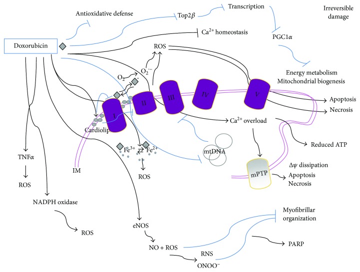
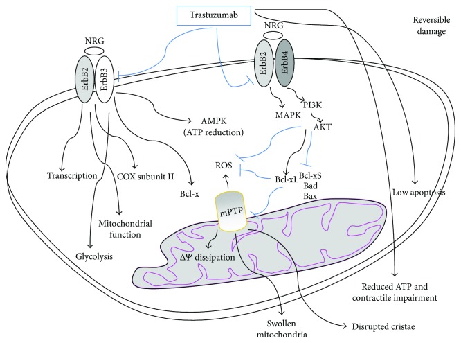
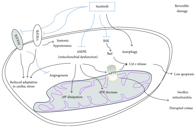
Many cancer therapies produce toxic side effects whose molecular mechanisms await full elucidation. The most feared and studied side effect of chemotherapeutic drugs is cardiotoxicity. Also, skeletal muscle physiology impairment has been recorded after many chemotherapeutical treatments. However, only doxorubicin has been extensively studied for its side effects on skeletal muscle. Chemotherapeutic-induced adverse side effects are, in many cases, mediated by mitochondrial damage. In particular, trastuzumab and sunitinib toxicity is mainly associated with mitochondria impairment and is mostly reversible. Vice versa, doxorubicin-induced toxicity not only includes mitochondria damage but can also lead to a more robust and extensive cell injury which is often irreversible and lethal. Drugs interfering with mitochondrial functionality determine the depletion of ATP reservoirs and lead to subsequent reversible contractile dysfunction. Mitochondrial damage includes the impairment of the respiratory chain and the loss of mitochondrial membrane potential with subsequent disruption of cellular energetic. In a context of increased stress, AMPK has a key role in maintaining energy homeostasis, and inhibition of the AMPK pathway is one of the proposed mechanisms possibly mediating mitochondrial toxicity due to chemotherapeutics. Therapies targeting and protecting cell metabolism and energy management might be useful tools in protecting muscular tissues against the toxicity induced by chemotherapeutic drugs.


Figures

Figure 1
Figure 1
Doxorubicin-mediated cytotoxicity is mostly irreversible. Mitochondrial doxorubicin accumulation is due to its specific binding to the phospholipid cardiolipin; this membrane perturbation inhibits complex I and complex II disrupting the electron transport chain and inducing ROS production. ROS might also be produced by other doxorubicin-mediated mechanisms: a quinone moiety in the chemical structure of doxorubicin is reduced by complex I into a reactive semiquinone free radical which transfers an electron to O2 and generates the superoxide anion O2•−. In turn, the semiquinone free radical is oxidized and returns to the quinone form in a sequence of reactions known as the “redox cycling” of doxorubicin. Moreover, doxorubicin can directly interact with iron to form reactive anthracycline-iron complexes resulting in an iron cycling between Fe3+ and Fe2+ associated with ROS production and altering iron homeostasis. Doxorubicin also induces mtDNA damage and binds to eNOS enhancing its activity thus leading to NO production and contributing to peroxynitrite (ONOO) formation. It also disrupts Ca2+ homeostasis which triggers mPTP and dissipates the transmembrane potential (ΔΨ) along with increasing mitochondrial permeability to apoptotic factors such as cytochrome c and leading to apoptosis or necrosis. The excessive oxidative stress produced by doxorubicin can also be mediated by increasing levels of TNFα and by NADPH oxidase and leads to redox modifications of macromolecules such as myofibrillar proteins. Doxorubicin also reduces the antioxidative defense of cells, and by preventing Top2β activity, it alters the transcriptome, for example, downregulating PGC-1α, which negatively impacts on both oxidative phosphorylation and mitochondrial biogenesis. IM: inner membrane space.
Figure 2
Figure 2
Trastuzumab-mediated cytotoxicity is mostly reversible. ErbB2 inhibition by trastuzumab reduces prosurvival signalings mediated by neuregulin-ErbB2-ErbB4 and is associated with a dramatic increase of proapoptotic Bcl-xS expression and a decrease of antiapoptotic Bcl-xL. This induces the opening of mPTP and generates ROS and mitochondrial dysfunctions among which loss of ΔΨ and ATP depletion with the disruption of cellular energetic, swollen mitochondria and reversibile contractile impairment. Cardiotoxicity of trastuzumab might also be related to its inhibition of AMPK which, in conditions of stress, leads to depletion of ATP stores. ErbB2/ErbB3 heterodimer controls Bcl-X and AMPK counteracting ATP depletion and destabilization of mitochondrial membrane, thus protecting contractile function. ErbB2 can translocate to the nucleus possibly acting on transcription and trastuzumab inhibition of ErbB2 has been suggested to regulate the expression of COX subunits.
Figure 3
Figure 3
Sunitinib-mediated cytotoxicity is mostly reversible. Sunitinib impinges on cellular energy homeostasis, via disrupting the mitochondrial function through the inhibition of AMPK signaling. This induces mPTP opening, ΔΨ dissipation, swollen mitochondria, disrupted cristae, and a massive decrease of intracellular ATP, but low cytochrome c release and apoptosis. Sunitinib has been suggested to be also able to increase the autophagic flux and to inhibit ribosomal protein S6 kinase (RSK), thus activating Bad. By inhibiting VEGFR and PDGFR, sunitinib impairs angiogenesis and reduces adaptation to cardiac stress.

References

    1. Waseem M., Parvez S. Mitochondrial dysfunction mediated cisplatin induced toxicity: modulatory role of curcumin. Food and Chemical Toxicology. 2013;53:334–342. doi: 10.1016/j.fct.2012.11.055. - DOI - PubMed
    1. Serrano J., Palmeira C. M., Kuehl D. W., Wallace K. B. Cardioselective and cumulative oxidation of mitochondrial DNA following subchronic doxorubicin administration. Biochimica et Biophysica Acta (BBA) - Bioenergetics. 1999;1411(1):201–205. doi: 10.1016/S0005-2728(99)00011-0. - DOI - PubMed
    1. Lauritzen K. H., Kleppa L., Aronsen J. M., et al. Impaired dynamics and function of mitochondria caused by mtDNA toxicity leads to heart failure. American Journal of Physiology-Heart and Circulatory Physiology. 2015;309(3):H434–H449. doi: 10.1152/ajpheart.00253.2014. - DOI - PubMed
    1. Lombardi A. A., Elrod J. W. mtDNA damage in the development of heart failure. American Journal of Physiology-Heart and Circulatory Physiology. 2015;309(3):H393–H395. doi: 10.1152/ajpheart.00475.2015. - DOI - PubMed
    1. Järvelä L. S., Kemppainen J., Niinikoski H., et al. Effects of a home-based exercise program on metabolic risk factors and fitness in long-term survivors of childhood acute lymphoblastic leukemia. Pediatric Blood & Cancer. 2012;59(1):155–160. doi: 10.1002/pbc.24049. - DOI - PubMed

LinkOut - more resources