Case Reports
. 2018 May 5;40(1):9.
doi: 10.1186/s40902-018-0147-3. eCollection 2018 Dec.

Anatomical repair of a bilateral Tessier No. 3 cleft by midfacial advancement

Affiliations
Case Reports

Anatomical repair of a bilateral Tessier No. 3 cleft by midfacial advancement

Ji-Hyeon Oh et al. Maxillofac Plast Reconstr Surg. .

Abstract

Background: Bilateral Tessier number 3 clefts are extremely rare, and their surgical treatments have not been well established.

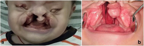
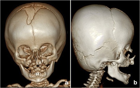
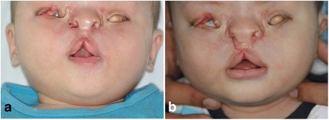
Case presentation: The authors describe the case of a patient with a right Tessier number 3, 11 facial cleft with microphthalmia, a left Tessier number 3 facial cleft with anophthalmia, and cleft palate. We repaired simultaneously the bilateral soft tissue clefts by premaxillary repositioning, cleft lip repair, facial cleft repair by nasal lengthening, midfacial advancement, and an upper eyelid transposition flap with repositioning both the medial canthi. Postoperatively, the patient showed an esthetically acceptable face without unnatural scars.

Conclusions: We achieved good results functionally and esthetically by midfacial advancement with facial muscle reposition instead of traditional interdigitating Z-plasties. The surgical modality of our anatomical repair and 3 months follow-up results are presented.

Keywords: Anatomic repair; Anophthalmos; Bilateral Tessier 3; Facial muscle reposition; Midfacial advancement.


Conflict of interest statement

Not applicable.Written informed consent was obtained from the patient’s guardian for publication of this case report and accompanying images.Both authors declare that they have no competing interests.Springer Nature remains neutral with regard to jurisdictional claims in published maps and institutional affiliations.

Figures

Fig. 1
Fig. 1
Preoperative clinical photographs. a An 8-month-old boy showed bilateral oronaso-ocular clefts with right-sided microphthalmia and left-sided clinical anophthalmia. b Intraorally, bilateral severe alveolar clefts and complete palatal cleft were revealed
Fig. 2
Fig. 2
3D CT views. a The frontal view demonstrated corresponding bony clefts extended from the alveolus, through the nasomaxillary sutures, and up to the inferomedial walls of the orbits. b The lateral view demonstrated a protruding premaxilla
Fig. 3
Fig. 3
Intraoperative photographs and the schematic design. a Incision lines were marked on the prolabium, alar bases, lateral lip segments, and peri-orbital area, after tattooing anatomical landmarks such as peak points for the Cupid’s bow, alar base, nasion, real and expected medial canthus, and mouth corners. b The schematic design of surgical incisions. c All the sutures are done
Fig. 4
Fig. 4
Postoperative follow-up photographs. a At suture removal. b 3 months after the operation

References

    1. Tessier P. Anatomical classification facial, cranio-facial and latero-facial clefts. J Maxillofac Surg. 1976;4:69–92. doi: 10.1016/S0301-0503(76)80013-6. - DOI - PubMed
    1. Eppley BL, van Aalst JA, Robey A, Havlik RJ, Sadove AM. The spectrum of orofacial clefting. Plast Reconstr Surg. 2005;115:101e–114e. doi: 10.1097/01.PRS.0000164494.45986.91. - DOI - PubMed
    1. Kalantar-Hormozi A, Abbaszadeh-Kasbi A, Goravanchi F, Davai NR. Prevalence of rare craniofacial clefts. J Craniofac Surg. 2017;28:e467–ee70. doi: 10.1097/SCS.0000000000003771. - DOI - PubMed
    1. Gawrych E, Janiszewska-Olszowska J, Chojnacka H. Tessier type 3 oblique facial cleft with a contralateral complete cleft lip and palate. Int J Oral Maxillofac Surg. 2010;39:1133–1136. doi: 10.1016/j.ijom.2010.05.012. - DOI - PubMed
    1. Wan DC, Lazareff JA, Jarrahy R, Bradley JP. Correction of large facial encephalocele with bilateral rare craniofacial clefts. J Craniofac Surg. 2011;22:338–342. doi: 10.1097/SCS.0b013e3181f7e0fb. - DOI - PubMed

Publication types