Review
. 2017 Jun 7;2(4):224-247.
doi: 10.1016/j.bioactmat.2017.05.007. eCollection 2017 Dec.

Bone grafts and biomaterials substitutes for bone defect repair: A review

Affiliations
Review

Bone grafts and biomaterials substitutes for bone defect repair: A review

Wenhao Wang et al. Bioact Mater. .

Abstract

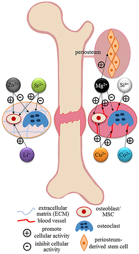
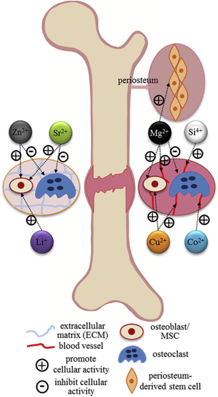
Bone grafts have been predominated used to treat bone defects, delayed union or non-union, and spinal fusion in orthopaedic clinically for a period of time, despite the emergency of synthetic bone graft substitutes. Nevertheless, the integration of allogeneic grafts and synthetic substitutes with host bone was found jeopardized in long-term follow-up studies. Hence, the enhancement of osteointegration of these grafts and substitutes with host bone is considerably important. To address this problem, addition of various growth factors, such as bone morphogenetic proteins (BMPs), parathyroid hormone (PTH) and platelet rich plasma (PRP), into structural allografts and synthetic substitutes have been considered. Although clinical applications of these factors have exhibited good bone formation, their further application was limited due to high cost and potential adverse side effects. Alternatively, bioinorganic ions such as magnesium, strontium and zinc are considered as alternative of osteogenic biological factors. Hence, this paper aims to review the currently available bone grafts and bone substitutes as well as the biological and bio-inorganic factors for the treatments of bone defect.

Keywords: Bioinorganic ions; Bone grafts and substitutes; Fracture healing; Growth factors.


Figures

Image 1
Graphical abstract
Fig. 1
Fig. 1
The hierarchical structure of bone ranging from microscale skeleton to nanoscale collagen and hydroxyapatite. Reprinted by permission from Macmillan Publishers Ltd: Nature Communication , copyright (2013).
Fig. 2
Fig. 2
Illustration of a typical fracture healing process, biological events, and cellular activities at different phases. The primary metabolic phases (blue bars) of fracture healing overlap with biological phases (brown bars). The time scale of healing is equivalent to a mouse closed femur fracture fixed with an intramedullary rod. Abbreviations: PMN, polymorphonuclear leukocyte. Reprinted by permission from Macmillan Publishers Ltd: Nature Reviews Rheumatology , copyright (2014).
Fig. 3
Fig. 3
Male patient (age 19 years old) with infected non-union after intramedullary nailing of an open tibial fracture. (A). Anteroposterior (AP) and lateral X-rays of the tibia illustrating osteolysis (white arrow) secondary to infection. The patient underwent removal of the nail, extensive debridement and a two-staged reconstruction of the bone defect, using the induced membrane technique for bone regeneration (the Masquelet technique). (B) Intraoperative pictures showing: (1) a 60 mm defect of the tibia (black arrow) at the second stage of the procedure; (2) adequate mechanical stability was provided with internal fixation (locking plate) bridging the defect, while the length was maintained (black arrow); (3) maximum biological stimulation was provided using autologous bone graft harvested from the femoral canal (black arrow, right), bone-marrow mesenchymal stem cells (broken arrow, left) and the osteoinductive factor bone morphogenetic protein-7 (center); (4) the graft was placed to fill the bone defect (black arrow). (C) Intraoperative fluoroscopic images showing the bone defect after fixation. (D) Postoperative AP and lateral X-rays at 3 months, showing the evolution of the bone regeneration process with satisfactory incorporation and mineralization of the graft (photographs courtesy of PVG) .
Fig. 4
Fig. 4
Most common specific targets of relevant bioinorganic ions in their role of therapeutic agents revealed by current researches .
Fig. 5
Fig. 5
Schematic diagram of hierarchical structure in bone and proposed mechanism of ion-exchange behavior. (a) Macroscopic bone. (b) Haversian osteons in cortical bone, consisting of several concentric lamellar layers that are built from parallel collagen fibers. (c) Fine structure of collagen fiber, consisting of collagen fibrils. (d) Collagen molecular packing with mineral in the fibril. Collagen molecules are shown as green and yellow rods. Mineral crystals are shown as blue tiles. (e) Single molecule triple helix. Reproduced with permission of the International Union of Crystallography .
Fig. 6
Fig. 6
Schematic of hypothesized intracellular signaling cascades by Mg ion stimulation of human bone mesenchymal stem cells (hBMSCs). Addition of MgSO4 will increase intracellular Mg concentration in undifferentiated hBMSCs. HIFs are then translocated into the cell nucleus and induce production of Collagen X-α1 and VEGF. In differentiated hBMSCs, Mg ion activates PGC-1a production, which induces the production of VEGF. Abbreviations: HIF, hypoxia-inducible factor; NFAT, nuclear factor of activated T-cells; PGC-1α, peroxisome proliferation-activated receptor gamma, coactivator 1α; ERRα, estrogen-related receptor α (Reprinted from Ref. , Copyright (2014), with permission from Elsevier).
Fig. 7
Fig. 7
Schematic diagram showing (A) diffusion of Mg2+ across the bone toward the periosteum that is innervated by DGR sensory neurons and enriched with PDSCs undergoing osteogenic differentiation into new bone. (B) The released Mg2+ enters DRG neurons via Mg2+ transporters or channels and promotes CGRP-vesicles accumulation and exocytosis. The DRG-released CGRP, in turn, activate the CGRP receptor in PDSCs, which triggers phosphorylation of CREB1 via cAMP and promotes the expression of genes contributing to osteogenic differentiation. Abbreviations: DGR, dorsal root ganglia; PDSCs, periosteum-derived stem cells; CREB1, cAMP-responsive element binding protein 1; cAMP: cyclic adenosine monophosphate (Reprinted by permission from Macmillan Publishers Ltd: Nature Medicine , copyright (2016)).
Fig. 8
Fig. 8
(A) A schematic showing the dual mechanism of action of strontium (Sr): the stimulatory role on bone-forming osteoblast cells and the inhibitory role on bone resorbing osteoclast cells. (B) A schematic showing how Sr activates osteoblastogenesis. Abbreviations: CaSR, calcium sensing receptor; ERK1/2, extracellular signal-regulated kinases 1/2; P38, a mitogen-activated protein kinases; PLC, phospholipase C; PKD, protein kinase D; PI3K, phosphatidylinositide 3-kinases; PKCβII, protein kinase C βII; NF-kB, nuclear factor kappa beta; NFATc, nuclear factors of activated T cells; PGE2, prostaglandin, E2; FGFR, fibroblast growth factor receptor (Reprinted from Refs. , , Copyright (2013, 2012), with permission from Elsevier).
Fig. 9
Fig. 9
Effect of silicate nanoplatelets on hMSCs differentiation. a) The addition of silicate nanoplatelets upregulate alkaline phosphatase (ALP) activity of hMSCs. b) The increase in the RUNX2 (green) and production of bone-related proteins such as osteocalcin (OCN, green), and osteopontin (OPN, red) was observed due to the addition of silicates. Cells in normal media without silicate particles act as a negative control whereas cells in osteoinductive medium serve as a positive control. Cell nuclei were counterstained with DAPI (blue). c) The protein production was quantified using image analysis from the fluorescence images. The intensity of protein per cell was quantified and later normalized by the control (hMSCs in normal growth media with no silicate particles) to obtain a fold-increase in the production of protein (Reprint with permission from Ref. [282]).

References

    1. Bhatt R.A., Rozental T.D. Bone graft substitutes. Hand Clin. 2012;28:457–468. - PubMed
    1. Greenwald A.S., Boden S.D., Goldberg V.M., Yaszemski M., Heim C.S. Bone-graft Substitutes: Facts, Fictions and Applications, AAOS 75th Annual Meeting. 2008. - PubMed
    1. Dimitriou R., Jones E., McGonagle D., Giannoudis P.V. Bone regeneration: current concepts and future directions. BMC Med. 2011;9:66. - PMC - PubMed
    1. Campana V., Milano G., Pagano E., Barba M., Cicione C., Salonna G., Lattanzi W., Logroscino G. Bone substitutes in orthopaedic surgery: from basic science to clinical practice. J. Mater. Sci. Mater. Med. 2014;25:2445–2461. - PMC - PubMed
    1. Bauer T.W., Muschler G.F. Bone graft materials: an overview of the basic science. Clin. Orthop. Relat. Res. 2000;371:10–27. - PubMed