. 2018 Sep;410(24):6141-6154.
doi: 10.1007/s00216-018-1106-7. Epub 2018 May 10.

Mussel-inspired 3D fiber scaffolds for heart-on-a-chip toxicity studies of engineered nanomaterials

Affiliations

Mussel-inspired 3D fiber scaffolds for heart-on-a-chip toxicity studies of engineered nanomaterials

Seungkuk Ahn et al. Anal Bioanal Chem. 2018 Sep.

Abstract

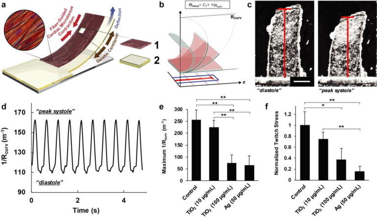
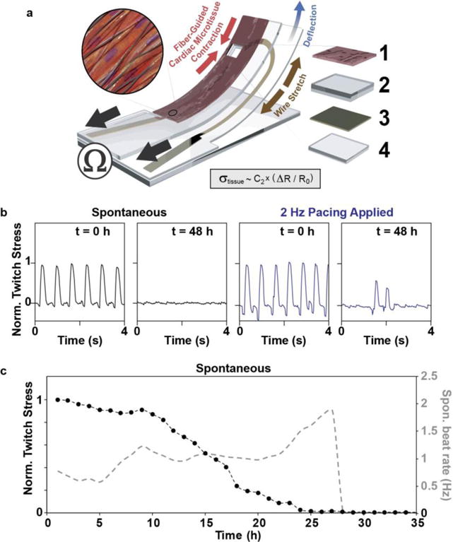
Due to the unique physicochemical properties exhibited by materials with nanoscale dimensions, there is currently a continuous increase in the number of engineered nanomaterials (ENMs) used in consumer goods. However, several reports associate ENM exposure to negative health outcomes such as cardiovascular diseases. Therefore, understanding the pathological consequences of ENM exposure represents an important challenge, requiring model systems that can provide mechanistic insights across different levels of ENM-based toxicity. To achieve this, we developed a mussel-inspired 3D microphysiological system (MPS) to measure cardiac contractility in the presence of ENMs. While multiple cardiac MPS have been reported as alternatives to in vivo testing, most systems only partially recapitulate the native extracellular matrix (ECM) structure. Here, we show how adhesive and aligned polydopamine (PDA)/polycaprolactone (PCL) nanofiber can be used to emulate the 3D native ECM environment of the myocardium. Such nanofiber scaffolds can support the formation of anisotropic and contractile muscular tissues. By integrating these fibers in a cardiac MPS, we assessed the effects of TiO2 and Ag nanoparticles on the contractile function of cardiac tissues. We found that these ENMs decrease the contractile function of cardiac tissues through structural damage to tissue architecture. Furthermore, the MPS with embedded sensors herein presents a way to non-invasively monitor the effects of ENM on cardiac tissue contractility at different time points. These results demonstrate the utility of our MPS as an analytical platform for understanding the functional impacts of ENMs while providing a biomimetic microenvironment to in vitro cardiac tissue samples. Graphical Abstract Heart-on-a-chip integrated with mussel-inspired fiber scaffolds for a high-throughput toxicological assessment of engineered nanomaterials.

Keywords: Cardiotoxicity; Microphysiological systems; Nanofiber; Nanotoxicology; Polydopamine.


Conflict of interest statement

a) Disclosure of potential conflicts of interest. The authors declare that they have no conflict of interest.

Figures

Figure 1
Figure 1
Nanofiber fabrication. (a) Schematic illustration of polymerization of PCL/DA into PCL/PDA through treatment with triethylammonium bicarbonate (TEAB, pH 8.5) buffer. (b–c) SEM images of b) PCL/DA and c) PCL/PDA. Insets indicate macroscopic images of nanofibers. Scales are 10 μm. d) Fiber diameter analysis. n=4. e) Fiber directionality calculated from (b–c) SEM images.
Figure 2
Figure 2
Physicochemical properties of nanofiber. a) FT-IR spectrum of PCL nanofiber, PCL/DA nanofiber, PCL/PDA film, and PCL/PDA nanofiber. b) Specific modulus of nanofiber scaffolds. n=4 for PCL/DA and n=5 for PCL/PDA. c–i) Adhesion of fibronectin on (c–e) PCL/PDA and (f–h) PCL/DA nanofiber. Scales are 100 μm. (i) FN coverages on the nanofiber were calculated and plotted. **p<0.05, n=3, ROI = at least 25 for each condition.
Figure 3
Figure 3
In vitro cardiomyocyte culture on nanofiber. a–d) (a–c) Bright field images of contraction of the fiber scaffolds with (d) kymograph. Scales of a-c are 100 μm. e) Confocal image of cardiomyocytes on nanofiber, stained for nuclei (blue) and α-actinin (grey). Scale is 20 μm. f–h) (f) Orientational order parameter (OOP), g) sarcomere length, and h) sarcomere packing density (SPD) of cardiomyocytes grown on the fibrous scaffolds. n=3 and ROI=4.
Figure 4
Figure 4
Measuring contractile stress using fiber-coated gelatin MPS. a) Principle sketch of a fiber-coated MPS with cardiomyocytes. Constituent layer 1: engineered cardiac tissue within nanofiber scaffolds and 2: gelatin thin film; see Electronic Supplementary Material Movie 2 for a representative video of a fiber-coated gelatin MPS seeded with contracting NRVMs. b) Schematic diagram showing the extrapolation of x-projections of cantilever deflection in 2D and its correlation to Rcurv. c) Optical images showing the cantilever motions associated to cardiomyocyte diastole and systole. Scale is 1 mm. d) Representative plot of the geometric readout from the optical recording of MPS motion over time, under 2 Hz pacing. e–f) Comparison of (e) maximum 1/Rcurv and (f) normalized twitch stress values calculated from Day 5 cardiac MPS samples under different ENM exposure conditions (48 h after ENM addition, 2 Hz pacing at 10 V), For statistical comparison, *p<0.10 and **p<0.05, n=10 for control, n=6 for 10 and 100 μg/ml and TiO2, and n=7 for Ag (50 μg/ml).
Figure 5
Figure 5
Fiber-coated cardiac microphysiological device with embedded contractility sensors. a) Principle sketch of device: an embedded flexible thin film sensor provides non-invasive electrical readout of contractile stress generated by PCL/PDA nanofiber-supported cardiac micro-tissue. Device adapted from Ref. [14] by introducing PCL/PDA nanofiber coating onto device surface. Constituent layer 1: engineered cardiac tissue within nanofiber scaffolds, 2: PDMS layer, 3: thin film sensor layer, and 4: bottom PDMS layer; see Electronic Supplementary Material Movie 3 for a representative video of a fiber-coated device seeded with contracting NRVMs. b) Electrical readout of normalized twitch stress generated by tissue prior to and after 48 hrs of exposure to (100 μg/ml) TiO2. c) Electrical readout of normalized twitch stress (black traces, left axis) and beat rate (mean (3) grey trace, right axis) generated spontaneously by tissue during first 35 h of exposure to (100 μg/ml) TiO2.
Figure 6
Figure 6
Effect of ENMs on calcium transient and sarcomere structure. a–h) Confocal images of calcium dye fluorescent (green) with calcium transient at the specific points (white boxes) for (a–b) control, (c–d) TiO2 (10 μg/ml) exposure, and (e–f) TiO2 (100 μg/ml) exposure. Scales are 500 μm. g–i) Confocal image of cardiomyocytes on nanofiber, stained for nuclei (blue) and α-actinin (grey). Scales are 100 μm (for the top panels) and 20 μm (for the bottom panels). The bottom panels are the zoom-in images from the red dots of the top panels. j) Orientational order parameter (OOP) analysis of cardiomyocytes after TiO2 exposure. For statistical comparison with respect to control, **p<0.05, n= 7 for control (0 μg/ml) and TiO2 (100 μg/ml) and 6 for TiO2 (10 μg/ml).

References

    1. Walser T, Limbach LK, Brogioli R, Erismann E, Flamigni L, Hattendorf B, et al. Persistence of engineered nanoparticles in a municipal solid-waste incineration plant. Nat Nanotechnol. 2012;7(8):520. - PubMed
    1. Simeonova PP, Erdely A. Engineered nanoparticle respiratory exposure and potential risks for cardiovascular toxicity: predictive tests and biomarkers. Inhalation Toxicol. 2009;21(sup1):68–73. - PubMed
    1. Yokel RA, MacPhail RC. Engineered nanomaterials: exposures, hazards, and risk prevention. J Occup Med Toxicol. 2011;6(1):7. - PMC - PubMed
    1. Gwinn MR, Vallyathan V. Nanoparticles: health effects—pros and cons. Environ Health Perspect. 2006;114(12):1818. - PMC - PubMed
    1. Stampfl A, Maier M, Radykewicz R, Reitmeir P, Göttlicher M, Niessner R. Langendorff heart: a model system to study cardiovascular effects of engineered nanoparticles. ACS nano. 2011;5(7):5345–53. - PubMed