

Pattern of muscle involvement in inclusion body myositis: a sonographic study
- PMID: 29745890
- PMCID: PMC6558657
Pattern of muscle involvement in inclusion body myositis: a sonographic study
Abstract
Objectives: Imaging plays a role in myositis assessment by detecting muscle changes indicative of pathology. This study was conducted to determine the ultrasonographic pattern of muscle involvement in patients with inclusion body myositis (IBM) through an assessment of muscle echointensity.
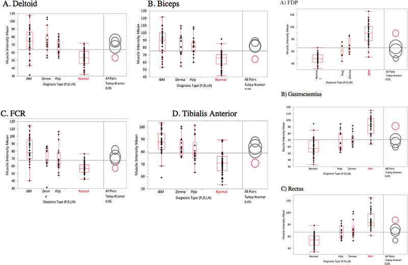
Methods: Sixty-two individuals were consecutively studied, 18 with IBM, 16 with polymyositis or dermatomyositis and 28 normal controls. Standardised scans were completed bilaterally for the deltoids, biceps, flexor digitorum profundus (FDP), flexor carpi ulnaris, rectus femoris, tibialis anterior and gastrocnemius assessing for muscle echointensity changes.
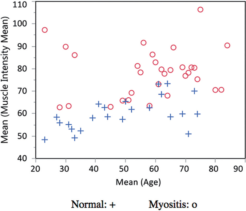
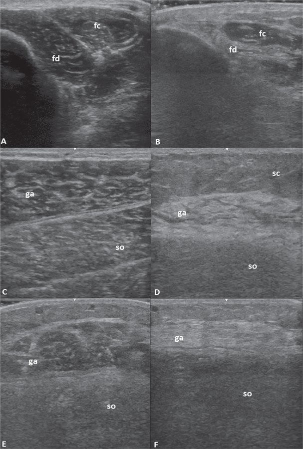
Results: Patients with IBM had a markedly increased muscle echointensity when compared with comparator groups for all muscles studied. This was most discriminating at the FDP, gastrocnemius and rectus femoris. Asymmetry between sides and a heterogeneously increased echointensity were also seen.
Conclusions: Ultrasonography can aid in the assessment of IBM by displaying an increased echointensity in characteristically involved muscles, particularly when combined with assessments for asymmetry and echotexture.
Figures
References
-
- NEEDHAM M, MASTAGLIA FL: Sporadic inclusion body myositis: A review of recent clinical advances and current approaches to diagnosis and treatment. Clin Neurophysiol 2016; 127: 1764–73. - PubMed
-
- HILTON-JONES D, BRADY S: Diagnostic criteria for inclusion body myositis. J Intern Med 2016; 280: 52–62. - PubMed
-
- TASCA G, MONFORTE M, DE FINO C et al.: Magnetic resonance imaging pattern recognition in sporadic inclusion-body myositis: Muscle MRI in IBM. Muscle Nerve 2015; 52: 956–62. - PubMed
-
- PHILLIPS BA, CALA LA, THICKBROOM GW et al.: Patterns of muscle involvement in inclusion body myositis: clinical and magnetic resonance imaging study. Muscle Nerve 2001; 24: 1526–34. - PubMed
-
- COX FM, REIJNIERSE M, VAN RIJSWIJK CSP et al.: Magnetic resonance imaging of skeletal muscles in sporadic inclusion body myositis. Rheumatology 2011; 50: 1153–61. - PubMed
Publication types
MeSH terms
Grants and funding
LinkOut - more resources
Full Text Sources