. 2018 Oct;32(10):5685-5702.
doi: 10.1096/fj.201701512RR. Epub 2018 May 10.

A lysosome-plasma membrane-sphingolipid axis linking lysosomal storage to cell growth arrest

Affiliations

A lysosome-plasma membrane-sphingolipid axis linking lysosomal storage to cell growth arrest

Maura Samarani et al. FASEB J. 2018 Oct.

Abstract

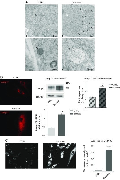
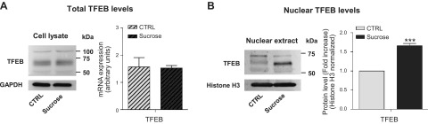
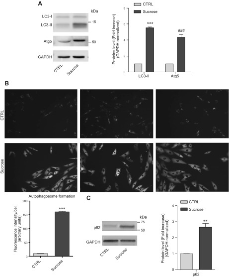
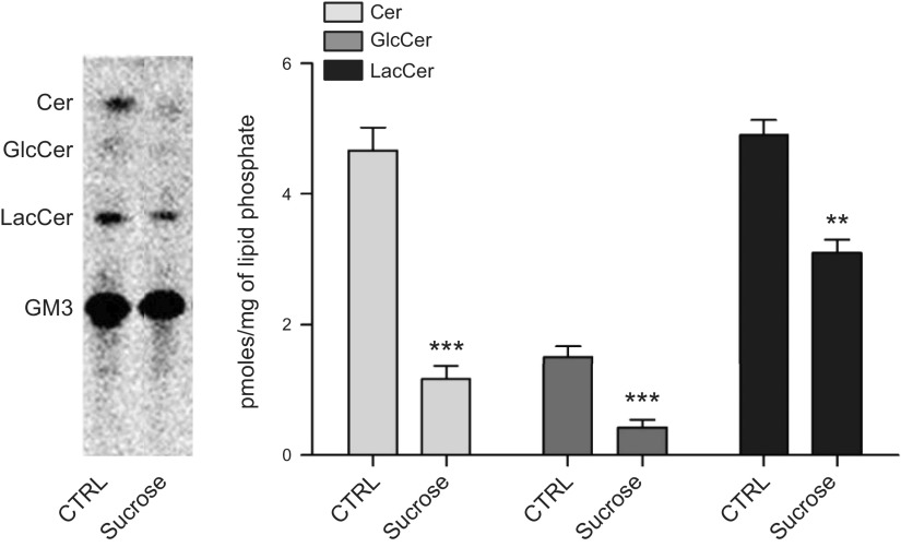
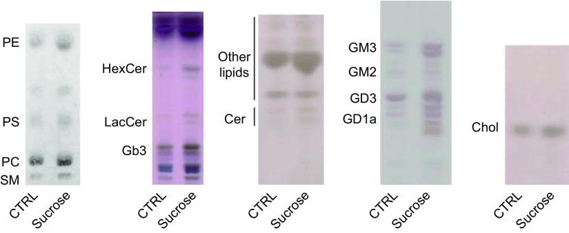
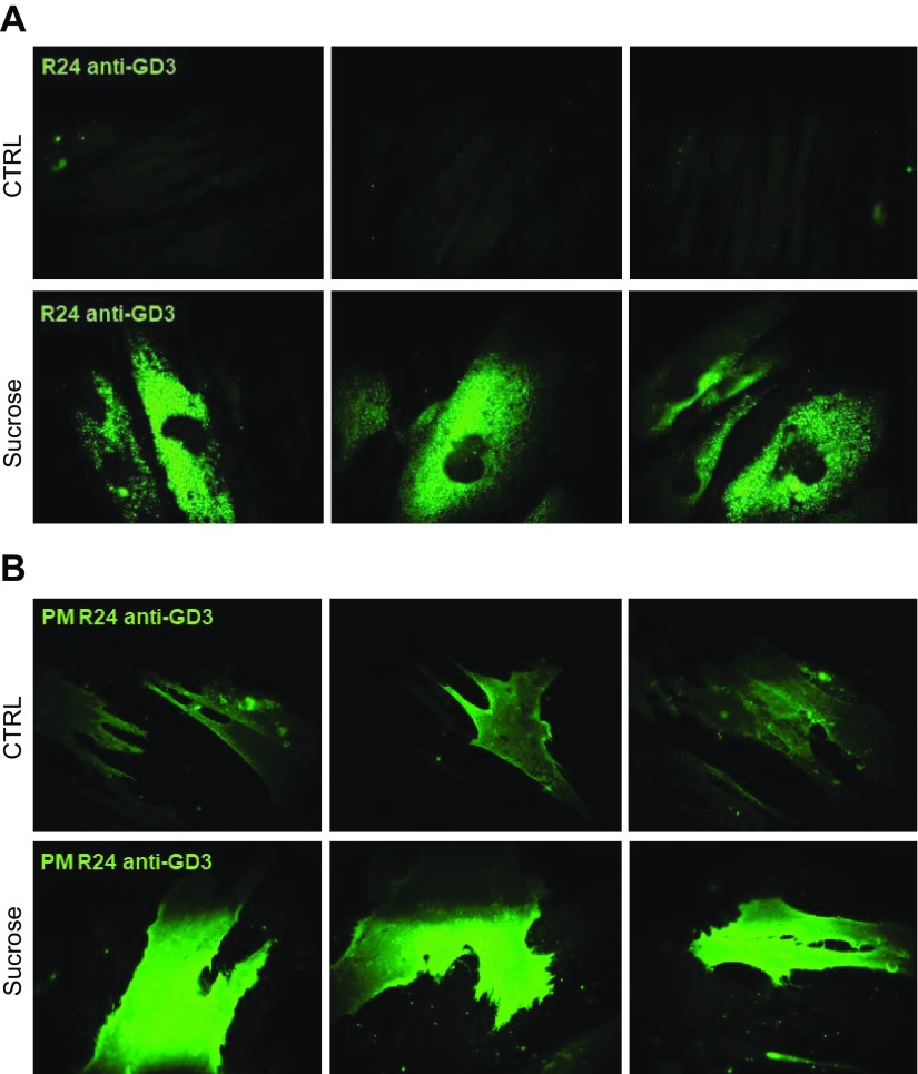
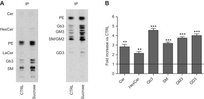
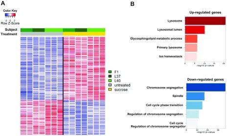
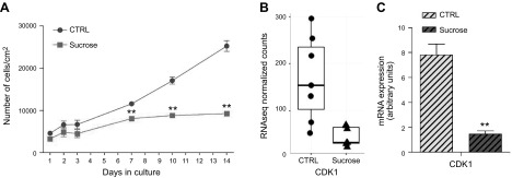
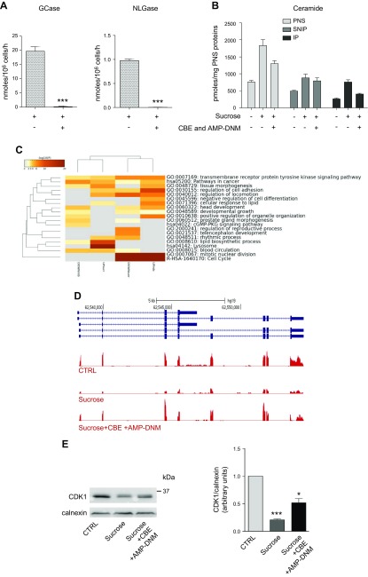
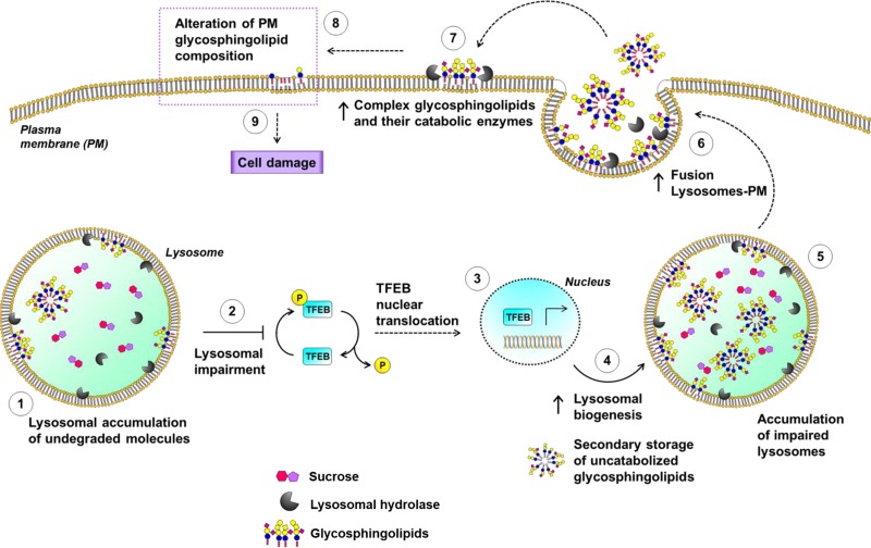
Lysosomal accumulation of undegraded materials is a common feature of lysosomal storage diseases, neurodegenerative disorders, and the aging process. To better understand the role of lysosomal storage in the onset of cell damage, we used human fibroblasts loaded with sucrose as a model of lysosomal accumulation. Sucrose-loaded fibroblasts displayed increased lysosomal biogenesis followed by arrested cell proliferation. Notably, we found that reduced lysosomal catabolism and autophagy impairment led to an increase in sphingolipids ( i.e., sphingomyelin, glucosylceramide, ceramide, and the gangliosides GM3 and GD3), at both intracellular and plasma membrane (PM) levels. In addition, we observed an increase in the lysosomal membrane protein Lamp-1 on the PM of sucrose-loaded fibroblasts and a greater release of the soluble lysosomal protein cathepsin D in their extracellular medium compared with controls. These results indicate increased fusion between lysosomes and the PM, as also suggested by the increased activity of lysosomal glycosphingolipid hydrolases on the PM of sucrose-loaded fibroblasts. The inhibition of β-glucocerebrosidase and nonlysosomal glucosylceramidase, both involved in ceramide production resulting from glycosphingolipid catabolism on the PM, partially restored cell proliferation. Our findings indicate the existence of a new molecular mechanism underlying cell damage triggered by lysosomal impairment.-Samarani, M., Loberto, N., Soldà, G., Straniero, L., Asselta, R., Duga, S., Lunghi, G., Zucca, F. A., Mauri, L., Ciampa, M. G., Schiumarini, D., Bassi, R., Giussani, P., Chiricozzi, E., Prinetti, A., Aureli, M., Sonnino, S. A lysosome-plasma membrane-sphingolipid axis linking lysosomal storage to cell growth arrest.

Keywords: catabolism; cell proliferation; cell surface; glycohydrolases; glycosphingolipids.


Conflict of interest statement

The authors thank Dr. Maria Carla Panzeri (Advanced Light and Electron Microscopy BioImaging Center, San Raffaele Scientific Institute, Milan, Italy) for her expert assistance in electron microscopy imaging, and Prof. J. M. F. G. Aerts (Leiden University, Leiden, The Netherlands) for providing AMP-DNM. This work was supported by Fondazione Cariplo Grant 2015–1017 to M.A., and was partially supported by the Italian National Research Council’s flagship “InterOmics” Project (PB.P05), part of the 2012–2014 program on aging sponsored by the National Research Program and the National Research Council (Italian Ministry of Education, Universities and Research). Lipidomic analyses were partially supported by the Medical University of South Carolina’s Lipidomics Shared Resource [Hollings Cancer Center, Medical University of South Carolina (MUSC); Grant P30 CA138313]; the Lipidomics Shared Resource of the South Carolina Lipidomics and Pathobiology Centers of Biomedical Research Excellence (COBRE); MUSC Department of Biochemistry (Grant P20 RR017677); and the U.S. National Institutes of Health, National Center for Research Resources and Office of the Director (Grant C06 RR018823) for the occupancy of space for the Lipidomics Shared Resource, 505 Children’s Research Institute (CRI) Analytical Unit. The authors declare no conflicts of interest.

Figures

Figure 1
Figure 1
Effect of sucrose loading on the endolysosomal compartment in human fibroblasts. A) Electron micrographs of human control (a, c) and sucrose-loaded fibroblasts (b, d). Arrowheads (b) and arrow (d) indicate sucrosomes; arrow (c) indicates lysosomes; N (a, b) indicates nucleus. B) Immunofluorescence staining of lysosomal marker Lamp-1 in permeabilized human fibroblasts, or not with sucrose (left). Immunostaining of Lamp-1 and loading control GAPDH accompanied with the semiquantitative graph of normalized Lamp-1/GAPDH (middle). Data are expressed as means ± sd. **P < 0.0021 vs. control (ctrl) (n = 4). Lamp-1 mRNA expression evaluated by quantitative RT-PCR (right). *P < 0.01 vs. ctrl (n = 3) Expression levels were normalized using HMBS and B2M as housekeeping genes. C) LysoTracker Red DND-99 staining performed on living human fibroblasts, loaded or not with sucrose, and a semiquantitative graph of the fluorescence intensity measured in ctrl and sucrose-loaded cells normalized by the number of cells analyzed. ***P < 0.0001 vs. ctrl (n = 70).
Figure 2
Figure 2
TFEB nuclear translocation. A) Immunostaining of TFEB from total cell lysates of control and sucrose-loaded cells (left). TFEB mRNA expression evaluated by quantitative RT-PCR (right). Expression levels were normalized using HMBS and B2M as housekeeping genes. Data are expressed as means ± sd; (n = 3). B) Immunostaining of TFEB in nuclear extracts from ctrl and sucrose-loaded cells (left). Semiquantitative graph of nuclear TFEB normalized as TFEB/histone H3 (right). ***P < 0.0003 vs. ctrl (n = 4).
Figure 3
Figure 3
Macroautophagic markers in sucrose-loaded cells. A) Immunostaining of LC3-I, LC3-II, and Atg5 along with a graph representing the fold change of protein levels over control (ctrl). Data are expressed as means ± sd. ***P < 0.0001 vs. ctrl, ###P < 0.0008 vs. ctrl (n = 3). B) Autophagosomes were evaluated in control and 14-d sucrose-loaded cells using an autophagosome detection assay. The pictures are representative of 3 experiments. The graph represents the fluorescence associated with each cell, data were obtained through the quantification of 70 cells for both sucrose-loaded and ctrl cells. ***P < 0.0001 (n = 3). C) Immunostaining of p62 accompanied by a graph representing the fold change of protein levels over ctrl. Data are expressed as means ± sd. **P < 0.0023 vs. ctrl (n = 3). GAPDH was used as the loading ctrl.
Figure 4
Figure 4
Sucrose-loaded fibroblasts are characterized by an impairment in SL degradation. Lysosomal catabolism of [3-3H(sphingosine)]GM3 in control and sucrose-loaded fibroblasts. Digital autoradiography of HPTLC (left); graph representing the quantification of GM3 catabolites (right). Data are expressed as picomoles of radioactive lipids per milligram of lipid phosphate. Cer, ceramide; GlcCer, glucosylceramide; LacCer, lactosylceramide. Data are expressed as means ± sd. ***P < 0.0005 vs. control (ctrl), **P < 0.005 vs. ctrl (n = 3).
Figure 5
Figure 5
Lipids levels in sucrose-loaded fibroblasts. HPTLC of phospholipids, neutral glycosphingolipids, ceramide, gangliosides, and cholesterol. Cer, ceramide; Chol, cholesterol; Gb3, globotriaosylceramide; HexCer, hexosylceramides; LacCer, lactosylceramide; PC, phosphatidylcholine; PE, phosphatidylethanolamine; PS, phosphatidylserine; SM, sphingomyelin.
Figure 6
Figure 6
Sucrose-loaded fibroblasts are characterized by an increased content of GD3 at both intracellular and PM levels. A) Indirect immunofluorescence of GD3 ganglioside obtained by staining with R24 antibody in human fibroblasts, loaded or not with sucrose permeabilized with Triton X-100. B) PM levels of GD3 ganglioside evaluated by indirect immunofluorescence in fibroblasts, loaded or not with sucrose with sucrose, in nonpermeabilizing conditions. Images are representative of 10 fields for each sample. Results represent 3 independent experiments performed in triplicate.
Figure 7
Figure 7
SL composition and content of detergent-resistant PM regions. A) Representative HPTLC of the radioactive lipids associated with the IP of PM biotinylated proteins performed with 2 solvent systems: chloroform:methanol:water [110:40:6 (v:v:v)] (a) , and chloroform:methanol:0.2% aqueous CaCl2 [50:42:11 (v:v:v)] (b). B) Graph representing the fold increase of each lipid in sucrose-loaded cells with respect to control (ctrl). ***P < 0.0002 vs. ctrl, **P < 0.004 vs. ctrl. The line represents the value fixed at 1 for the ctrl.
Figure 8
Figure 8
Lysosomal accumulation impacts on the expression of lysosome- and cell cycle–related genes. A) Heatmap showing selective gene expression alterations in fibroblasts loaded with sucrose for 14 d (3 biological replicates—L37, L40, F1—each performed in duplicate or triplicate; n = 14). Each row was normalized by z score. B) GO analysis showing enrichment of functional terms among sucrose-induced (red) and sucrose-repressed (blue) genes.
Figure 9
Figure 9
Aberrant catabolism of complex glycosphingolipids on the PM. A) Immunofluorescence of PM-associated Lamp-1 in human fibroblasts loaded or not with sucrose. B) Immunostaining of Lamp-1 associated with immunoprecipitated PM biotinylated proteins (biotin-IP) from sucrose-loaded and control cells. C) Released cathepsin D in culture medium from control (ctrl) and sucrose-loaded cells after 14 d, detected by ELISA. ***P < 0.0002 vs. ctrl. D) PM-associated activities of GCase, NLGase, β-galactosidase, and β-hexosaminidase. Activities were normalized to nmol/106 cells/h. Data are expressed as means ± sd. ***P < 0.0007 vs. ctrl (n = 4). E) HPTLC of [3-3H(sphingosine)]GM3 catabolites produced at the PM level in ctrl and sucrose-loaded fibroblasts. The graph represents the quantification of GM3 catabolites. Data are expressed as mean ± sd percentages of radioactivity with respect to the total radioactivity associated with the lipid extracts. ***P < 0.0005 vs. ctrl, **P < 0.005 vs. ctrl. Cer, ceramide; GlcCer, glucosylceramide; LacCer, lactosylceramide.
Figure 10
Figure 10
Cell growth arrest in sucrose-loaded cells. A) Cell growth curve of control and sucrose-loaded fibroblasts on d 1–3, 7, 10, and 14. Data are expressed as number of living cells per square centimeter of growth area. **P < 0.0094. B) CDK1 expression evaluated by RNAseq on fibroblasts from 3 individuals (n = 7), FDR-corrected P = 0.86 × 10⁻⁵ (left). CDK1 mRNA expression evaluated by qPCR. Expression levels were normalized using HMBS and B2M as housekeeping genes (right). **P < 0.0022 (n = 3).
Figure 11
Figure 11
Cell growth arrest is rescued by the inhibition of glycosphingolipid catabolism on the PM. A) PM-associated activities of GCase and NLGase in sucrose-loaded cells treated or untreated with GCase and NLGase inhibitors (CBE and AMP-DNM, respectively). Activities were normalized to nmol/106 cells/h ± sd. ***P < 0.0005 vs. sucrose (n = 3). B) Graph representing the picomoles of ceramide associated with the fraction obtained by streptavidin separation after cell-surface biotinylation from PNS preparation in conditions that preserved the lipid–protein interactions. Bars refer to control, sucrose-loaded cells, and sucrose-loaded cells treated with inhibitors. *P < 0.04 vs. ctrl, **P < 0.006 vs. ctrl, #P < 0.03 vs. sucrose, ##P < 0.009 vs. sucrose (n = 2). Data are expressed as pmol/mg PNS protein. C) Comparison of GO enrichment analysis among DE genes in sucrose-loaded cells untreated or treated with GCase and NLGase inhibitors after 14 d. The heatmap reports P values for significance of enrichment; gray cells indicate a lack of enrichment. D) Snapshot of RNAseq expression data for the CDK1 gene, showing down-regulated and enhanced mRNA levels, respectively, in fibroblasts treated with sucrose alone or with sucrose and GCase and NLGase inhibitors (CBE + AMP-DNM). E) Immunostaining of CDK1 evaluated in control (ctrl), 14-d sucrose-loaded cells (sucrose), and 14-d sucrose-loaded cells treated with GCase and NLGase inhibitors (sucrose + CBE + AMP-DNM); calnexin was used as the loading ctrl (left). A semiquantitative graph of normalized CDK1/calnexin (right). ***P < 0.0001 vs. ctrl, *P < 0.02 vs. sucrose (n = 3).
Figure 12
Figure 12
A suggested molecular mechanism linking lysosomal engulfment to the onset of cell damage: crosstalk between lysosomes and PM.

References

    1. Appelmans F., Wattiaux R., De Duve C. (1955) Tissue fractionation studies. 5. The association of acid phosphatase with a special class of cytoplasmic granules in rat liver. Biochem. J. 59, 438–445 - PMC - PubMed
    1. De Duve C., Pressman B. C., Gianetto R., Wattiaux R., Appelmans F. (1955) Tissue fractionation studies. 6. Intracellular distribution patterns of enzymes in rat-liver tissue. Biochem. J. 60, 604–617 - PMC - PubMed
    1. Saftig P., Klumperman J. (2009) Lysosome biogenesis and lysosomal membrane proteins: trafficking meets function. Nat. Rev. Mol. Cell Biol. 10, 623–635 - PubMed
    1. Perera R. M., Zoncu R. (2016) The lysosome as a regulatory hub. Annu. Rev. Cell Dev. Biol. 32, 223–253 - PMC - PubMed
    1. Platt F. M., Boland B., van der Spoel A. C. (2012) The cell biology of disease: lysosomal storage disorders: the cellular impact of lysosomal dysfunction. J. Cell Biol. 199, 723–734 - PMC - PubMed

Publication types