. 2018:599:21-68.
doi: 10.1016/bs.mie.2017.11.035. Epub 2018 Jan 10.

Fe-S Clusters and MutY Base Excision Repair Glycosylases: Purification, Kinetics, and DNA Affinity Measurements

Affiliations

Fe-S Clusters and MutY Base Excision Repair Glycosylases: Purification, Kinetics, and DNA Affinity Measurements

Nicole N Nuñez et al. Methods Enzymol. 2018.

Abstract

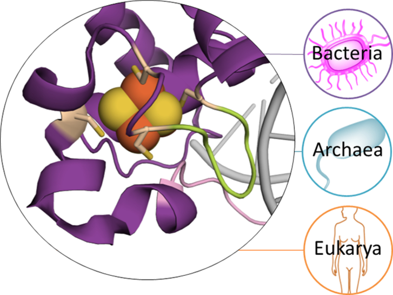
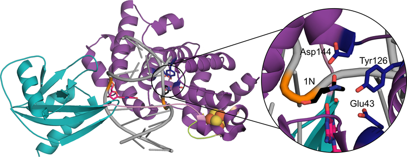
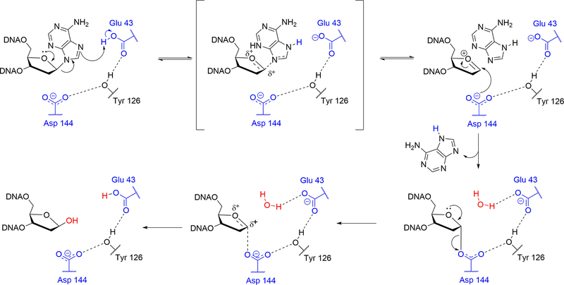
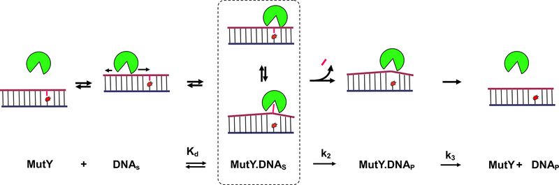
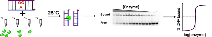
A growing number of iron-sulfur (Fe-S) cluster cofactors have been identified in DNA repair proteins. MutY and its homologs are base excision repair (BER) glycosylases that prevent mutations associated with the common oxidation product of guanine (G), 8-oxo-7,8-dihydroguanine (OG) by catalyzing adenine (A) base excision from inappropriately formed OG:A mispairs. The finding of an [4Fe-4S]2+ cluster cofactor in MutY, Endonuclease III, and structurally similar BER enzymes was surprising and initially thought to represent an example of a purely structural role for the cofactor. However, in the two decades subsequent to the initial discovery, purification and in vitro analysis of bacterial MutYs and mammalian homologs, such as human MUTYH and mouse Mutyh, have demonstrated that proper Fe-S cluster coordination is required for OG:A substrate recognition and adenine excision. In addition, the Fe-S cluster in MutY has been shown to be capable of redox chemistry in the presence of DNA. The work in our laboratory aimed at addressing the importance of the MutY Fe-S cluster has involved a battery of approaches, with the overarching hypothesis that understanding the role(s) of the Fe-S cluster is intimately associated with understanding the biological and chemical properties of MutY and its unique damaged DNA substrate as a whole. In this chapter, we focus on methods of enzyme expression and purification, detailed enzyme kinetics, and DNA affinity assays. The methods described herein have not only been leveraged to provide insight into the roles of the MutY Fe-S cluster but have also been provided crucial information needed to delineate the impact of inherited variants of the human homolog MUTYH associated with a colorectal cancer syndrome known as MUTYH-associated polyposis or MAP. Notably, many MAP-associated variants have been found adjacent to the Fe-S cluster further underscoring the intimate relationship between the cofactor, MUTYH-mediated DNA repair, and disease.

Keywords: DNA damage; DNA repair; DNA–protein interactions; Enzyme kinetics; Fe–S cluster; Protein purification.


Figures

Figure 1.
Figure 1.. MutY Fe-S Cluster.
As seen in the crystal structure of Geobacillus stearothermophilus MutY (PDB 5DPK) (purple) (Woods et al., 2016), the [4Fe-4S]2+ cluster (orange and yellow spheres) is coordinated by four Cys ligands (beige colored sticks), and homolog lineages bearing the iconic Fe-S cluster span throughout all three domains of life. Notably, the Fe-S cluster is important for providing key contacts with the DNA (grey) via positioning of the FCL (green) generated by the first two Cys in the Cys-X6-Cys-X2-Cys-X5-Cys conserved motif, and is located adjacent to the start of the IDC (light pink) (Guan et al., 1998).
Figure 2.
Figure 2.. Structure and Updated Mechanism for MutY
a) Crystal structure of Gs MutY (PDB ID: 5DPK) bound to DNA (grey) containing OG (hot pink) opposite the pyrrolidine transition state mimic, 1N (black) (Woods et al., 2016). MutY has several structural features, including the N-terminal domain (purple) that houses the Fe-S cluster (orange and yellow spheres) and FCL (green), which is connected to the C-terminal domain (teal), via the IDC (light pink). b) Mechanism of adenine removal by MutY and homologs where active site residues are marked in blue and numbered according to their positions in Gs MutY. The adenine base is first protonated by Glu43 to form a good leaving group, and dissociates from the sugar via an SN1-like mechanism. The resulting oxocarbenium ion is proposed to be stabilized though the formation of a covalent intermediate with Asp144, which dissociates upon nucleophilic attack by an activated water molecule (red) to form the abasic site product.
Figure 3.
Figure 3.. Representative chromatogram of Mutyh during purification through a Hi-Trip Heparin column.
The blue, red and pink lines refer to the absorbance at 280 nm, 260 nm and 410 nm wavelengths, respectively, while the green and brown lines refer to the percentage of heparin buffer B and the conductivity of the sample. Mutyh and its homologs have similar chromatograms, and typically elute at a NaCl concentration of 450 mM as shown by the peak above. The peak eluted just prior to the main protein peak is truncated protein and is collected in separate fractions to prevent contamination of the full-length enzyme (Pope et al., 2005).
Figure 4.
Figure 4.. MutY Glycosylase Assays.
Schematic depiction of the glycosylase assay and PAGE for determination of kinetic parameters and active fractions of MutY/MUTYH.
Figure 5.
Figure 5.. Schematic representation of damage processing and minimal kinetics scheme for MutY and homologs.
The free enzyme is known to non-specifically engage with the DNA strand, and use a processive search to detect the OG:A lesion. Upon encountering the lesion, it flips the adenine into its active site pocket. The binding affinity of the enzyme-substrate complex is determined by the dissociation constant Kd. The enzyme then catalyzes base excision (measured by the rate constant k2) to form an abasic site product, which it then slowly releases (measured by rate constant k3).
Figure 6.
Figure 6.. General Set-up for glycosylase assays
a) Set-up for a multiple-turnover/active site titration experiment showing composition of the reaction and quench tubes. b) Representative gel from a typical active site experiment. c) Characteristic plot of the concentration of product produced over time. Note, single-turnover experiments would be performed similarly by adjusting the enzyme concentration to be in excess.
Figure 7.
Figure 7.. EMSA for determination of Kd.
Schematic illustration of assay set up, representative gel image, and expected plot of gel quantitation of Kd curve.
Figure 8.
Figure 8.. EMSA Determination of koff.
Schematic of assay set up, representative gel of separation of enzyme bound and unbound DNA, and graphing of the image results. The first lane in gel is the negative control, containing no enzyme, second lane is positive control with no unlabeled DNA added.
Figure 9.
Figure 9.. MAP variants located at Pro281 (cyan) and Arg295 (magenta) are adjacent the Fe-S cluster.
The MUTYH N-terminal (purple) fragment crystal structure of Hs MUTYH (PDB 3N5N) (Luncsford et al., 2010) contains the FCL (light green), IDC (light pink) and Fe-S cluster (orange and yellow spheres) that is coordinated by four cysteine residues (beige colored sticks)

References

    1. Al-Tassan N, Chmiel NH, Maynard J, Fleming N, Livingston AL, Williams GT, Hodges AK, Davies DR, David SS, Sampson JR, and Cheadle JR (2002). Inherited variants of MYH associated with somatic G : C -> T : A mutations in colorectal tumors. Nat Genet 30, 227–232. - PubMed
    1. Au KG, Cabrera M, Miller JH, and Modrich P (1988). Escherichia coli mutY gene product is required for specific A-G----C.G mismatch correction. Proc Natl Acad Sci U S A 85, 9163–9166. - PMC - PubMed
    1. Au KG, Clark S, Miller JH, and Modrich P (1989). Escherichia coli mutY gene encodes an adenine glycosylase active on G-A mispairs. Proceedings of the National Academy of Sciences 86, 8877–8881. - PMC - PubMed
    1. Bai H, Jones S, Guan X, Wilson TM, Sampson JR, Cheadle JP, and Lu A-L (2005). Functional characterization of two human MutY homolog (hMYH) missense mutations (R227W and V232F) that lie within the putative hMSH6 binding domain and are associated with hMYH polyposis. Nucleic Acids Res 33, 597–604. - PMC - PubMed
    1. Banda DM, Nuñez NN, Burnside MA, Bradshaw KM, and David SS (2017). Repair of 8- oxoG:A mismatches by the MUTYH glycosylase: Mechanism, metals and medicine. Free Radical Biology and Medicine 107, 202–215. - PMC - PubMed

Publication types

MeSH terms